- Reduced price

Order to parcel locker

easy pay

 Delivery policy
Choose Paczkomat Inpost, Orlen Paczka, DHL, DPD or Poczta Polska. Click for more details
 Security policy
Pay with a quick bank transfer, payment card or cash on delivery. Click for more details
 Return policy
If you are a consumer, you can return the goods within 14 days. Click for more details
Interpreting X-ray images correctly is essential for diagnostic radiographers, as well as a widely used skill for emergency department doctors, nurse practitioners, and many other healthcare professions. This new title provides a systematic, methodical approach to musculoskeletal image interpretation and its role in the evaluation and treatment of injury.
A companion to the eighth edition of Bones and Joints, this book covers the basic principles for interpreting images and then follows a simple regional approach to common radiographic projections. It goes on to consider common and important fracture patterns and other injuries related to that region, as well as the differences between normal and abnormal images.
Image Interpretation is an ideal learning guide for undergraduates, those transitioning to graduate roles or clinical practice, and other healthcare professionals wanting to supplement their training.
Data sheet
1. Principles of Image Interpretation
2. Normal appearances of bones and joints
3. Fractures and Joint Trauma
4. Upper limb
5. Shoulder girdle and thorax
6. Lower limb
7. Pelvic girdle
8. Spine
9. Facial bones and mandible
10. Practical applications of image interpretation
Reference: 90420
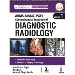
Author: Manavjit Singh Sandhu
Reference: 93560
Author: Michael N. Patlas
General Principles
Reference: 93586
Author: Loren Ketai