- Obniżka

Medycyna estetyczna

Diagnoza i psychoterapia uzależnień

Zamawiaj do paczkomatu

Płać wygodnie

 Dostawa
Wybierz Paczkomat Inpost, Orlen Paczkę, DHL, DPD, Pocztę, email (dla ebooków). Kliknij po więcej
 Płatność
Zapłać szybkim przelewem, kartą płatniczą lub za pobraniem. Kliknij po więcej szczegółów
 Zwroty
Jeżeli jesteś konsumentem możesz zwrócić towar w ciągu 14 dni*. Kliknij po więcej szczegółów
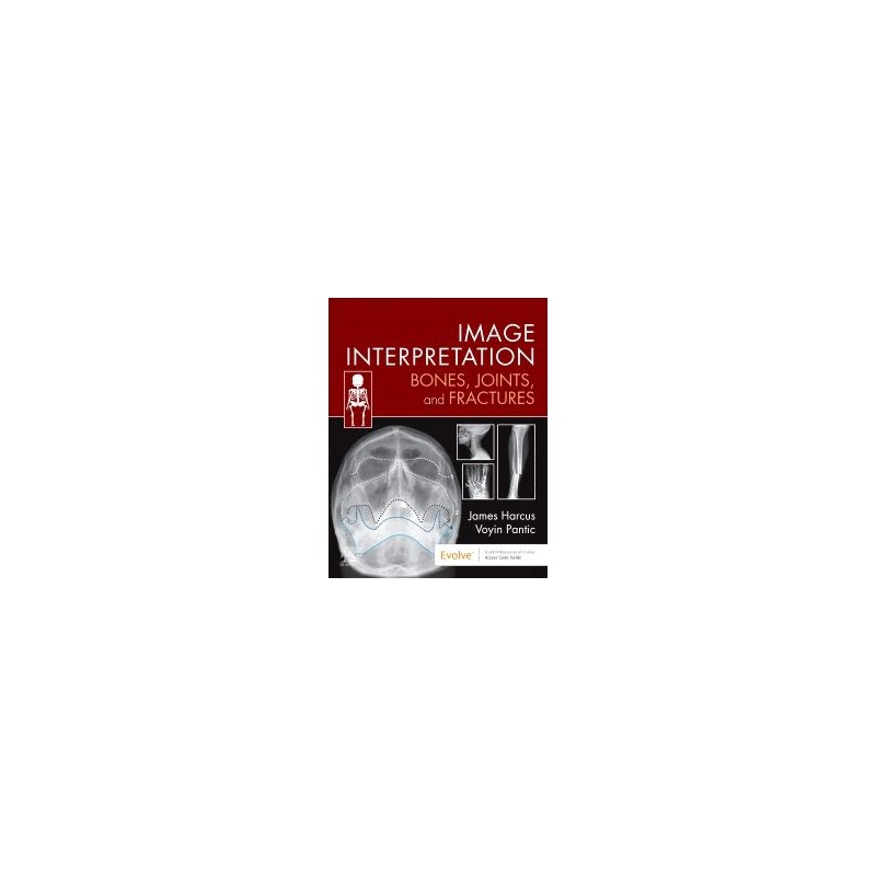
Interpreting X-ray images correctly is essential for diagnostic radiographers, as well as a widely used skill for emergency department doctors, nurse practitioners, and many other healthcare professions. This new title provides a systematic, methodical approach to musculoskeletal image interpretation and its role in the evaluation and treatment of injury.
A companion to the eighth edition of Bones and Joints, this book covers the basic principles for interpreting images and then follows a simple regional approach to common radiographic projections. It goes on to consider common and important fracture patterns and other injuries related to that region, as well as the differences between normal and abnormal images.
Image Interpretation is an ideal learning guide for undergraduates, those transitioning to graduate roles or clinical practice, and other healthcare professionals wanting to supplement their training.
Opis
1. Principles of Image Interpretation
2. Normal appearances of bones and joints
3. Fractures and Joint Trauma
4. Upper limb
5. Shoulder girdle and thorax
6. Lower limb
7. Pelvic girdle
8. Spine
9. Facial bones and mandible
10. Practical applications of image interpretation
Indeks: 90420
Autor: Manavjit Singh Sandhu
Indeks: 93586
Autor: Loren Ketai