- Reduced price

Order to parcel locker

easy pay

 Delivery policy
Choose Paczkomat Inpost, Orlen Paczka, DPD or Poczta Polska. Click for more details
 Security policy
Pay with a quick bank transfer, payment card or cash on delivery. Click for more details
 Return policy
If you are a consumer, you can return the goods within 14 days. Click for more details
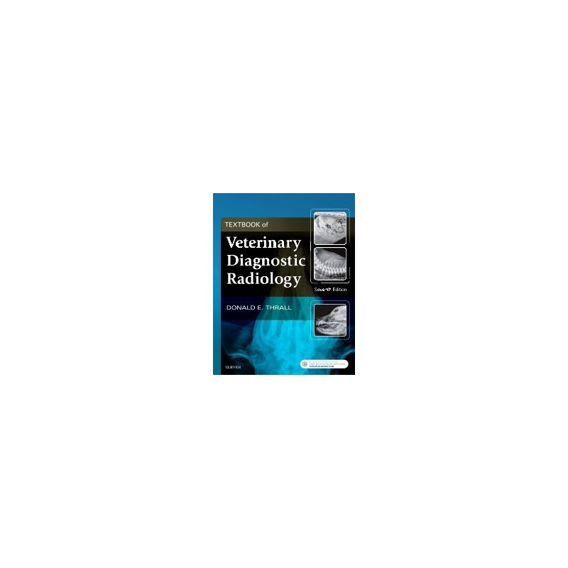
**Selected for Doody’s Core Titles® 2024 with Essential Purchase designation in Veterinary Medicine**
Learn the latest advances in veterinary diagnostic radiology! Textbook of Veterinary Diagnostic Radiology, 7th Edition, is a one-stop resource covering the principles of radiographic technique and interpretation for dogs, cats, and horses. Within this bestselling text, high-quality radiographic images accompany clear coverage of diagnostic radiology, ultrasound, MRI, and CT. User-friendly direction helps you to develop essential skills in patient positioning, radiographic technique and safety measures, normal and abnormal anatomy, radiographic viewing and interpretation, and alternative imaging modalities. This new edition has been thoroughly revised to include important advances in the field, information about contrast media, dental radiography, and more!
Data sheet
Section I: Physics and Principles of Interpretation
1. Radiation Protection and Physics of Diagnostic Radiology
2. Digital Radiographic Imaging
3. Physics of Ultrasound Imaging
4. Physics of Computed Tomography and Magnetic Resonance Imaging
5. Radiographic, CT and MR Contrast Media
6. Introduction to Radiographic Interpretation
Section II: The Axial Skeleton: Canine, Feline, and Equine
7. Radiographic Anatomy of the Axial Skeleton
8. Basic Principles of Radiographic Interpretation of the Axial Skeleton
9. The Cranial and Nasal Cavities: Canine and Feline
10. Magnetic Resonance Imaging Features of Brain Disease in Small Animals
11. The Equine Head
12. The Canine and Feline Vertebrae
13. Magnetic Resonance Imaging and Computed Tomography Features of Canine and Feline Spinal Cord Disease
Section III: The Appendicular Skeleton: Canine, Feline, and Equine
14. Radiographic Anatomy of the Appendicular Skeleton
15. Principles of Radiographic Interpretation of the Appendicular Skeleton
16. Orthopedic Diseases of Young and Growing Dogs and Cats
17. Fracture Healing and Complications
18. Radiographic Features of Bone Tumors and Bone Infections
19. Radiographic Signs of Joint Disease in Dogs and Cats
20. The Equine Stifle and Tarsus
21. The Equine Carpus
22. The Equine Metacarpal and Metatarsal Regions
23. The Equine Metacarpophalangeal and Metatarsophalangeal Articulation
24. The Equine Phalanges
25. The Equine Navicular Bone
Section IV: Thoracic Cavity: Canine, Feline, and Equine
26. Principles of Radiographic Interpretation of the Thorax
27. The Pharnyx, Larynx, and Trachea
28. Canine and Feline Esophagus
29. The Thoracic Wall
30. The Diaphragm
31. The Mediastinum
32. The Pleural Space
33. The Heart and Pulmonary Vessels
34. The Canine and Feline Lung
35. The Equine Thorax
Section V: Abdominal Cavity: Canine and Feline
36. Principles of Radiographic Interpretation of the Abdomen
37. The Peritoneal Space
38. The Liver and Spleen
39. The Kidneys and Ureters
40. The Urinary Bladder
41. The Urethra
42. The Prostate Gland
43. The Uterus, Ovaries and Testes
44. The Stomach
45. The Small Bowel
46. The Large Bowel
Reference: 71250
Author: Lois Hickman
Reference: 5481
Author: Robert Wood
Reference: 9449
Author: Jerzy Ziętek
z elementami zoonoz, wybranymi zagadnieniami z hodowli, anatomii i fizjologii
Reference: 69047
Author: Dennis C. Tanner
Reference: 79826
Author: Andrew Holloway