- Reduced price

Order to parcel locker

easy pay


 Delivery policy
Choose Paczkomat Inpost, Orlen Paczka, DPD or Poczta Polska. Click for more details
 Security policy
Pay with a quick bank transfer, payment card or cash on delivery. Click for more details
 Return policy
If you are a consumer, you can return the goods within 14 days. Click for more details
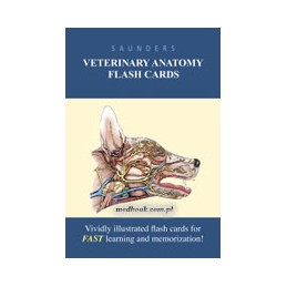
Study anywhere, anytime and master veterinary anatomy with Saunders Veterinary Anatomy Flash Cards. Included in this set of 360 flash cards are approximately 400 full-color illustrations. The front of the card shows the anatomical drawing with numbered lead lines pointing to different anatomic structures, allowing you to quiz yourself on identification. The numeric answer key on the back of the card provides an easy comprehension check.
Data sheet
Introduction: How to Use This Book
Part 1: The Head and Ventral Neck
Canine Anatomy
1-1 Tympanic Bullae and Petrous Temporal Bones
1-2 Lymph Nodes and Lymphatic Vessels
1-3 Skull (lateral view)
1-4 Skull (dorsal view)
1-5 Skull (ventral view)
1-6 Skull (lateral view)
1-7 Mandible
1-8 Hyoid Apparatus and Larynx
1-9 Hard and Soft Palate
1-10 Salivary Glands
1-11 Simple Tooth
1-12 Esophagus
1-13 Head at the Level of P2
1-14 Head at the Level of the Eyeball
1-15 Thyroid Gland
1-16 Arteries of the Head
1-17 Closure of the Neural Plate
1-18 Gray Substance of Spinal Cord and Medulla Oblongata
1-19 Spinal Cord
1-20 Brain (ventral view)
1-21 Cerebral Hemispheres
1-22 Brain (lateral view)
1-23 Cortical Lobes of the Brain
1-24 Visceral and Somatic Nervous System
1-25 Meninges of the Brain
1-26 Production and Circulation of Cerebrospinal Fluid
1-27 Trigeminal Nerve
1-28 Autonomic Innervation of Structures of the Head
1-29 Parasympathetic Nervous System
1-30 Sympathetic Nervous System
1-31 Three Tunics of the Eye
1-32 Retina
1-33 Stumps of Ocular Muscles
1-34 Principal Arteries Supplying the Eye
1-35 Tympanic Bullae and Petrous Temporal Bones
1-36 Left Auricular Cartilage
1-37 Paramedian Section of the Head
1-38 Head, Rostral Part of the Orbit
1-39 Salivary Glands
1-40 Disarticulated Puppy Skull
1-41 CT Image of Caudal Nasal Cavity
1-42 Superficial Branches of Facial and Trigeminal Nerves
1-43 Branches of the Common Carotid Artery
1-44 Muscles, Nerves, and Salivary Glands
1-45 Cranial Nerves Leaving the Skull
1-46 Meninges and Ventricles of Brain, Median Plane
1-47 Sulci of Brain
1-48 Gyri of Brain
1-49 Brain, Cranial Nerves, and Brain Stem
Feline Anatomy
1-50 Tomogram of the Nasal Cavity
1-51 Ear Canal and Middle Ear
Equine Anatomy
1-52 Superficial Muscles of the Head
1-53 Permanent Dentition
1-54 Paramedian Section of the Head
1-55 Head at the Level of P4
1-56 Laryngeal Cartilages
1-57 Intrinsic Muscles of the Larynx
1-58 Hypophysis
1-59 Superficial Dissection of the Head
1-60 Deeper Dissection of the Head
1-61 Head at the Level of the Rostral Maxillary
1-62 CT Scan (Bone Window) of Head at Level of the Rostral Maxillary
1-63 Median Section of the Head
1-64 Topography of Conchofrontal and Maxillary Sinuses
1-65 Brain and Frontal and Maxillary Sinuses
1-66 Tongue and Pharynx
1-67 Root Convergence of Permanent Lower Incisors
1-68 Nasopharynx and Larynx
1-69 Deep Dissection of the Head
1-70 Dissection of the Orbit
1-71 Principal Arteries of the Head
1-72 Lymphatic Structures of the Head and Neck
Ruminant Anatomy
1-73 Superficial Dissection of the Head
1-74 Skull
1-75 Skin Innervation of the Head
1-76 Paranasal Sinuses
1-77 Neck
1-78 Lymph Drainage of Head and Neck
Porcine Anatomy
1-79 Head of a 9-month-old
1-80 Lymph Centers of the Head and Neck
1-81 Dissection of the Neck to Show Lymph Nodes
Avian Anatomy
1-82 Skull of a Chicken
Part 2: The Neck, Back, and Vertebral Column
Canine Anatomy
2-1 Structure of a Lymph Node
2-2 Cervical Vertebrae
2-3 Thoracic Vertebrae
2-4 Sacrum and Caudal Vertebrae
2-5 Trunk Muscles, Deeper Layers
2-6 Trunk Muscles, Deepest Layers
2-7 Thoracic and Lumbar Vertebrae
2-8 Epaxial Muscles
2-9 Epaxial Muscles
2-10 Superficial Nerves of the Neck
2-11 Veins of the Neck
2-12 Peripheral Distribution of Sympathetic and Parasympathetic Divisions
Feline Anatomy
2-13 Thoracic and Lumbar Vertebrae
Equine Anatomy
2-14 Nuchal Ligament and Associated Bursae
Ruminant Anatomy
2-15 Lumbar Intervertebral Disc
2-16 Vertebral Canal
Avian Anatomy
2-17 Oropharynx Opened by the Reflection of the Lower Jaw
Part 3: The Thorax
Canine Anatomy
3-1 Left Rib
3-2 Sternum and Costal Cartilages
3-3 Pleura and Pericardium
3-4 Thoracic Organs (left side)
3-5 Thoracic Organs (right side)
3-6 Lungs and Partial Aorta
3-7 Transformation of the Aortic Arches
3-8 Pericardium
3-9 Right Ventricle
3-10 Cardiac Nerves and Related Ganglia
3-11 Developing Heart
3-12 Vasa Vasorum
3-13 Branching of the Aortic Arch
3-14 Arteries of the Female Pelvis
3-15 Lymph Node
3-16 Palpable Lymph Nodes
3-17 Thoracic Lymph Nodes
3-18 Trunk
3-19 Vessels on the Floor of the Thorax
3-20 Heart and Lungs (left surface projection)
3-21 Heart and Lungs (right surface projection)
3-22 Right lung (lateral bronchogram)
3-23 Right lung (ventrodorsal bronchogram)
3-24 Trunk
3-25 Thoracic Cavity (left lateral view)
3-26 Thoracic Cavity (right lateral view)
3-27 Trunk (tranverse section at 7th thoracic vertebra)
3-28 Heart (lateral view)
3-29 Heart (ventrodorsal view)
3-30 Arteries of Thorax
Feline Anatomy
3-31 Thoracic, Abdominal, and Pelvic Cavities
3-32 Palpable Lymph Nodes
Equine Anatomy
3-33 Structures within the Mediastinum
3-34 Thorax at the Level of T5
3-35 Thorax at the Level of the Atrioventricular Valves
Ruminant Anatomy
3-36 Heart (left view)
3-37 Heart (right view)
3-38 Trunk at the Eighth Thoracic Vertebra
3-39 Lymph Drainage, Thoracic Wall and Mediastinum
Porcine Anatomy
3-40 Adult Lung
3-41 Seven- to Eight-Somite Embryo
3-42 Lungs
Avian Anatomy
3-43 Skeleton of a Chicken
3-44 Opened Syrinx
Part 4: The Abdomen
Canine Anatomy
4-1 Abdomen
4-2 Abdominal Cavity
4-3 Interior of the Stomach
4-4 Celiac Artery
4-5 Male Perineal Region
4-6 Cranial and Caudal Mesenteric Arteries
4-7 Formation of the Portal Vein
4-8 Bile Drainage System
4-9 Developing Simple Stomach
4-10 Development of the Pancreas
4-11 Growth and Rotation of the Midgut (stage one, lateral view)
4-12 Growth and Rotation of the Midgut (stage two, lateral view)
4-13 Growth and Rotation of the Midgut (stage three, lateral view)
4-14 Termination of the Abdominal Aorta
4-15 Abdominal Viscera
4-16 Abdominal Walls
4-17 Abdomen (lateral view)
4-18 Abdomen (ventrodorsal view)
4-19 Blood Supply of Stomach and Spleen
4-20 Abdomen after Administration of Barium Suspension (lateral view)
4-21 Abdomen after Administration of Barium Suspension (ventrodorsal view)
4-22 Abdomen at the Level of the First Lumbar Vertebra
4-23 Abdomen at Level of the First Lumbar Vertebra
4-24 Abdomen After Administration of a Barium Suspension (ventrodorsal view)
4-25 Trunk (dorsal section at level of the kidneys)
4-26 Abdomen at the Level of the Seventh Lumbar Vertebra
4-27 CT Image of Abdomen at the Level of the Seventh Lumbar Vertebra
4-28 Rectus Sheath
4-29 Peritoneal Reflections, Sagittal Section
4-30 Peritoneal Schema, Transverse Section through the Epiploic Foramen
Feline Anatomy
4-31 Abdomen After Administration of a Barium Suspension (lateral view)
4-32 Abdomen After Administration of a Barium Suspension (ventrodorsal view)
Equine Anatomy
4-33 Structures of the Abdominal Floor
4-34 Visceral Projections: Left Abdominal Wall
4-35 Visceral Projections: Right Abdominal Wall
4-36 Cecum and Related Organs
4-37 Visceral Projections: Ventral Abdominal Wall
4-38 Major Arteries of the GI Tract
4-39 Abdominal Autonomic Nerves and Branches of Abdominal Aorta
Ruminant Anatomy
4-40 Nerves to the Flank and Udder
4-41 Stomach
4-42 Greater and Lesser Omenta
4-43 Trunk at 13th Thoracic Vertebra
4-44 Disposition of the Greater Omentum
4-45 Abdominal Viscera of a Newborn Calf
4-46 Abdominal Viscera of a 5-Year-Old Cow
4-47 Abdominal Viscera of a6-Year-Old Pregnant Cow
4-48 Visceral Surface of the Liver
4-49 Reticulum of the Goat
4-50 Rumen of the Goat
Porcine Anatomy
4-51 Liver
4-52 Large Intestine
4-53 Visceral Surface of the Liver
4-54 Major Abdominal Arteries and Lymph Nodes
4-55 Lymph Nodes of the Sublumbar Area
Avian Anatomy
4-56 Stomach and Duodenum Loop
Part 5: The Pelvis and Reproductive Organs
Canine Anatomy
5-1 Embryos Showing Lateral Mesoderm and Celom
5-2 Intermediate Mesoderm
5-3 Development of the Testis
5-4 Urogenital Sinus
5-5 Development of the Ovary
5-6 Formation of the Vagina
5-7 Peritoneal Disposition
5-8 Testis and Epididymis
5-9 Canine Testis
5-10 Corrosion Cast of the Deferent Duct
5-11 Accessory Reproductive Glands
5-12 Functional Changes in the Female Tract
5-13 Ovary and the Ovarian Bursa
5-14 Formation of Extraembryonic Membranes
5-15 Topography of Adrenal Glands
5-16 Development of the Skin
5-17 Pelvis (transverse section at the level of the hip joint)
5-18 Reproductive Organs of the Bitch
5-19 Bladder, Urethra, and Penis
5-20 Quiescent and Erect Penis
Feline Anatomy
5-21 Bladder
Equine Anatomy
5-22 Bony Pelvis and Sacrosciatie Ligament
5-23 Pelvis of the Mare
5-24 Disposition of the Periotneum in the Pelvis
5-25 Sections of Equine Ovaries in Various Functional States
5-26 31-Day Twin Embryos
5-27 Pelvic Urethra and Accessory Reproductive Glands
5-28 Abdominal Pelvic Cavities
Ruminant Anatomy
5-29 Paraxial Mesoderm in an Embryo
5-30 Bony Pelvis
5-31 Bovine Pelvis at the Hip Joint
5-32 Blood Supply to the Reproductive Tract
5-33 Pelvis and Related Urogenital Organs of a Bull
5-34 Udder, Abdominal Floor and Cranial Quarters
5-35 Udder, Pelvic Floor and Caudal Quarters
Porcine Anatomy
5-36 Principal Arteries, Left Side of Female Reproductive Tract
5-37 Reproductive Organs of the Boar
Avian Anatomy
5-38 Male Reproductive Organs
5-39 Reproductive Organs of a Female Chicken
5-40 Fertilized Egg
Part 6: The Forelimb
Canine Anatomy
6-1 Synovial Joints
6-2 Transection of a Skeletal Muscle
6-3 Architecture of Skeletal Muscles
6-4 Ventral Muscles of the Neck and Thorax
6-5 Left Scapula
6-6 Left Humerus
6-7 Left Ulna and Radius
6-8 Carpal Skeleton
6-9 Right Manus
6-10 Superficial Muscles of the Shoulder and Arm
6-11 Intrinsic Muscles of the Left Shoulder and Arm
6-12 Muscles of the Left Forearm
6-13 Arteries of the Forelimb
6-14 Footpads of Fore- and Hindlimbs
6-15 Left Forelimb (transverse section at the level of the scapula)
6-16 Shoulder Joints (lateral and craniocaudal views)
6-17 Left Forelimb (transverse section distal to the elbow joint)
6-18 Arteries of the Right Forelimb
6-19 Elbow Joint (lateral view)
6-20 Elbow Joint (craniocaudal view)
6-21 Autonomous Zones of the Cutaneous Innervation of the Forelimb
6-22 Left Forelimb and Flexor Surfaces of the Joints
6-23 Left Forelimb
6-24 Capsule of the Left Shoulder Joint
6-25 Distribution of Musculocutaneous and Median Nerves, Right Forelimb
6-26 Distribution of Radial Nerve, Right Forelimb
6-27 Distribution of Ulnar Nerve: Right Forelimb
6-28 Veins of the Neck, Thoracic Inlet, and Proximal Forelimb
6-29 Blood Supply and Innervation Digits
Feline Anatomy
6-30 Claw
6-31 Autonomous Zones of the Cutaneous Innervation of the Forelimb
Equine Anatomy
6-32 Carpal Skeleton
6-33 Synovial Joint
6-34 Joints: Flexion, Extension, and Overextension
6-35 Muscles: Ventral Surface of the Thorax
6-36 Deep Muscles Attaching Forelimb to the Trunk
6-37 Left Forelimb
6-38 Shoulder Joint
6-39 Muscles: Medial Surface of Right Shoulder and Arm
6-40 Elbow Joint (lateral view)
6-41 Synovial Structures of the Left Shoulder and Elbow
6-42 Carpus (dorsopalmar radiograph)
6-43 Carpus (lateral radiograph)
6-44 Synovial Structures of the Left Carpus
6-45 Synovial Structures of the Left Carpus
6-46 Right Forearm
6-47 Structures Supporting the Fetlock Joint
6-48 Fetlock Joint and Digit
6-49 Axial Section of Digit
6-50 Ground Surface of the Hoof
6-51 Section of the Hoof
6-52 Transverse Section of the Hoof
6-53 Enlargement of the Dermal Lamellae
6-54 Dermis Exposed by Removal of Hoof
6-55 Major Arteries of Right Forelimb (medial view)
6-56 Medial Palmar Nerve
Ruminant Anatomy
6-57 Humerus
6-58 Carpal Skeleton
6-59 Muscles of the Forelimb
6-60 Foot (dorsopalmar radiograph)
6-61 Foot (lateromedial radiograph)
6-62 Forefoot (palmar view)
6-63 Right Forefoot (dorsal view)
6-64 Forefoot, Ground Surface of the Hoofs
6-65 Principal Arteries of Right Forelimb
6-66 Principal Veins of the Right Forelimb
6-67 Nerves of the Forelimb
6-68 Principal Nerves of the Right Forefoot
Porcine Anatomy
6-69 Carpal Skeleton
6-70 Phylogenetic Development: Horn Structures Associated with the Distal Phalanx
6-71 Palmar Surface of the Foot
Avian Anatomy
6-72 Skeleton of the Left Wing
6-73 Superficial Dissection of the Laterally Extended Left Wing
Part 7: The Hindlimb
Canine Anatomy
7-1 Hip Bone (lateral view)
7-2 Hip Bone (ventral view)
7-3 Sacrotuberous Ligament
7-4 Left Femur
7-5 Left Tibia and Fibula
7-6 Bones of the Tarsal Skeleton
7-7 Right Pes
7-8 Left Stifle Joint
7-9 Left Stifle Joint Showing Extent of Joint Capsule
7-10 Left Stifle Joint with the Patella Removed
7-11 Muscles of the Hindquarter and Thigh
7-12 Muscles of the Left Leg
7-13 Arteries of the Hindlimb
7-14 Pelvis with Extended and Flexed Hip Joints
7-15 Stifle (lateral view)
7-16 Left leg
7-17 Arteries of the Right Hindlimb
7-18 Hocks and Hindpaws
7-19 Hock
7-20 Cutaneous Innervation of the Hindlimb
7-21 Bones of the Left Pelvic Limb
7-22 Muscle Attachments on the Pelvis and Left Pelvic Limb
7-23 Arteries of Right Pelvic Limb
7-24 Veins of the Right Pelvic Limb
7-25 Distribution of Saphenous, Femoral, and Obturator Nerves of Right Pelvic Limb
7-26 Cranial and Caudal Gluteal Nerves and Sciatic Nerve of Right Pelvic Limb
Feline Anatomy
7-27 Pelvis (ventrodorsal and lateral views)
7-28 Stifle (craniocaudal view)
Equine Anatomy
7-29 Bones of the Tarsal Skeleton
7-30 Muscles of the Croup and Thigh
7-31 Croup Muscles, Resected to Expose the Ischial Tuber
7-32 Left Stifle Joint
7-33 Sagittal Section of Hock Joint
7-34 Stifle and Leg
7-35 Principal Arteries of Right Hindlimb
7-36 Branching of the Aortic Arch
7-37 Nerves of Right Hindfoot
Ruminant Anatomy
7-38 Bones of the Tarsal Skeleton
7-39 Muscles of the Left Hindlimb
7-40 Left Stifle Joint
7-41 Principal Arteries of the Right Hindlimb
7-42 Major Veins of the Hindlimb
7-43 Lymph Nodes of the Pelvis and Hindlimb
7-44 Nerves of the Right Hindlimb
Porcine Anatomy
7-45 Bones of the Tarsal Skeleton
7-46 Lymph Flow of the Hindlimb
