- Reduced price

Order to parcel locker

easy pay


 Delivery policy
Choose Paczkomat Inpost, Orlen Paczka, DHL, DPD or Poczta Polska. Click for more details
 Security policy
Pay with a quick bank transfer, payment card or cash on delivery. Click for more details
 Return policy
If you are a consumer, you can return the goods within 14 days. Click for more details
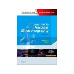
Now in its 6th edition, Introduction to Vascular Ultrasonography, by Drs. John Pellerito and Joseph Polak, provides an easily accessible, concise overview of arterial and venous ultrasound. A new co-editor and new contributors have updated this classic with cutting-edge diagnostic procedures as well as new chapters on evaluating organ transplants, screening for vascular disease, correlative imaging, and more. High-quality images, videos, and online access make this an ideal introduction to this complex and rapidly evolving technique.
Data sheet
SECTION 1: BASICS
SECTION 2: CEREBRAL VESSELS
The role of ultrasound in the management of cerebrovascular disease Normal cerbrovascular anatomy and collateral pathways Normal findings and technical aspects of carotid sonography Ultrasound assessment of carotid plaque Ultrasound assessment of carotid stenosis Carotid occlusion, unusual carotid pathology and tricky carotid cases Ultrasound assessment of the vertebral arteries Ultrasound assessment of the intracranial arteriesSECTION 3: EXTREMITY ARTERIES
Arterial anatomy of the extremities Nonimaging physiologic tests for assessment of lower extremity arterial disease Assessment of upper extremity arterial occlusive arterial disease Ultrasound evaluation before and after hemodialysis access Ultrasound assessment of lower extremity arteries Ultrasound assessment during and after carotid peripheral intervention Ultrasound in the assessment and management of arterial emergenciesSECTION 4: EXTREMITY VEINS
Risk factors and the role of ultrasound in the management of extremity venous disease Extremity venous anatomy and technique for ultrasound examination Ultrasound diagnosis of lower extremity venous thrombosis Controversies in venous ultrasound Ultrasound diagnosis of venous insufficiency Nonvascular pathology encountered during venous sonographySECTION 5: ABDOMEN AND PELVIS
Anatomy and normal doppler signatures of abdominal vessels Ultrasound assessment of the abdominal aorta Ultrasound imaging assessment following endovascular aortic aneurysm repair Ultrasound assessment of the splanchnic (mesenteric) arteries Ultrasound assessment of the hepatic vasculature Ultrasound assessment of native renal vessels Duplex ultrasound evaluation of the uterus and ovaries Duplex ultrasound evaluation of the male genitalia Evaluation of Organ Transplants Screening for Vascular Disease Correlative imaging Accreditation and the vascular labReference: 99300
Author: G. Richard Scott
Reference: 98865
Author: Irina Pollard
Reference: 11647
Author: Wojciech Braksator
