• Order to parcel locker

    Order to parcel locker
  • easy pay

    easy pay
  • Reduced price
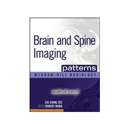
Brain and Spine Imaging Patterns

Brain and Spine Imaging Patterns

9780071465410
1,406.10 zł
1,335.80 zł Save 70.30 zł Tax included
Lowest price within 30 days before promotion: 1,335.80 zł
Quantity
Available in 4-6 weeks

  Delivery policy

Choose Paczkomat Inpost, Orlen Paczka, DPD or Poczta Polska. Click for more details

  Security policy

Pay with a quick bank transfer, payment card or cash on delivery. Click for more details

  Return policy

If you are a consumer, you can return the goods within 14 days. Click for more details

Description

Publishers Note:: Products purchased from Third Party sellers are not guaranteed by the publisher for quality, authenticity, or access to any online entitlements included with the product.


Sharpen your diagnostic skills for brain and spine disorders with this unique patterns-based approach to learning

Brain and Spine Imaging Patterns presents a systematic approach to understanding one of the most challenging areas of radiologic interpretation. Uniquely organized by various patterns seen on CT, MRI, and plain radiography imaging rather than pathology, the book carefully guides you toward a group of differential diagnoses. You will find an unmatched collection of more than 140 patterns covering:: skull defects and lesions; meningeal and sulcal diseases; extracerebral masses; intracerebral masses; mass lesions in the region of the ventricular system; sellar and parasellar masses; vascular legions; lesions in the cortical gray matter, white matter, and deep gray matter; and spinal diseases and lesions.

The easy-to-navigate organization of this book is specifically designed for use at the workstation. The concise text, numerous images, and helpful icons facilitate access to essential information and simplify the learning process.

Features

  • More than 140 patterns and more than 2500 digital-quality images
  • A strong focus on patterns recognized on MRI, including contrast-enhanced MRI
  • Icons, a grading system depicting the relative frequency of findings from common to rare, and the consistent organization of chapters help to clarify information for at-the-bench consultation
  • Many patterns include a set of “Related Patterns” images, which serve as cross-references to similar patterns for a given disorder
  • Special emphasis on the latest diagnostic modalities includes state-of-the-art depiction of image findings
Product Details
45541
9780071465410
9780071465410

Data sheet

Publication date
2010
Issue number
1
Cover
hard cover
Pages count
768
Dimensions (mm)
224 x 282
Weight (g)
2450
  • Contributors
    Foreword
    Preface
    Acknowledgments
    PART I: MAGNETIC RESONANCE IMAGING
    Section 1: Brain
    1. Skull Defects and Lesions
    2. Meningeal and Sulcal Diseases
    3. Extracerebral Masses
    4. Intracerebral Masses
    A. Sypratentorial Intraaxial Masses
    B. Infratentorial Intraaxial Masses
    5. Mass Lesions in the Region of the Ventricular System
    6. Sellar and Parasellar Masses
    7. Vascular Lesions
    8. Lesions in the Cortical Gray Matter, White Matter, and Deep Gray Matter
    Section 2: Spine
    9. Spinal Disease
    10. Extradural Disease
    11. Intradural Extramedullary Lesions
    12. Intramedullary Lesions
    PART II: COMPUTED TOMOGRAPHY
    Section 1: Brain
    13. Skull Defects and Lesions
    14. Meningeal and Sulcal Diseases
    15. Extracerebral Masses
    16. Intracerebral Masses
    A. Supratentorial Intraaxial Masses
    B. Infratentorial Intraaxial Masses
    17. Mass Lesions in the Region of the Ventricular System
    18. Sellar and Parasellar Masses
    19. Vascular Lesions
    20. Lesions in the Cortical Gray Matter, White Matter, and Deep Gray Matter
    Section 2: Spine
    21. Spinal Disease
    22. Extradural Lesions
    23. Intradural Extramedullary Lesions
    PART III: PLAIN RADIOGRAPHY
    24. Skull Defects and Lesions
    Index
Comments (0)