- Reduced price

Order to parcel locker

easy pay


 Delivery policy
Choose Paczkomat Inpost, Orlen Paczka, DHL, DPD or Poczta Polska. Click for more details
 Security policy
Pay with a quick bank transfer, payment card or cash on delivery. Click for more details
 Return policy
If you are a consumer, you can return the goods within 14 days. Click for more details
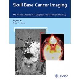
Skull base anatomy is extremely complex, with vital neurovascular structures passing through multiple channels and foramina. Brain tumors such as pituitary tumors, acoustic neuromas, and meningiomas are challenging to treat due to their close proximity to cranial nerves and blood vessels in the brain, neck, and spinal cord. Medical imaging is an essential tool for identifying lesions and critical adjacent structures. Detecting and precisely mapping out the extent of disease is imperative for appropriate and optimal treatment planning and ultimately patient outcome.
Eugene Yu and Reza Forghani have produced an exceptional, imaging-focused guide on various neoplastic diseases affecting the skull base, with contributions from a Whos Who of prominent radiologists, head and neck surgeons, neurosurgeons, and radiation oncologists. The content is presented in a clear and concise fashion with chapters organized anatomically. From the Anterior Cranial Fossa, Nasal Cavity, and Paranasal Sinuses - to the Petroclival and Lateral Skull Base, an overview and detailed analysis is provided for each region.
Key Highlights
This invaluable resource chronicles current knowledge in state-of-the-art skull base tumor imaging with clinical pearls on pathophysiology, prognosis, and treatment options. It is a must-have for radiology, neurosurgery, and otolaryngology residents and clinicians who care for patients with head and neck neoplasms.
Data sheet
1 Anterior Cranial Fossa, Nasal Cavity, and Paranasal Sinuses
2 Sellar, Parasellar, and Clival Region
3 Cerebellopontine Angle and Jugular Fossa
4 Petroclival and Lateral Skull Base
5 Open and Endoscopic Approaches to the Sinonasal Cavity and Skull Base
6 Posttreatment Appearance Following Skull Base Therapy
7 Neuroendovascular Procedures for Skull Base Neoplasia
8 Cross-Sectional Computed Tomography and Magnetic Resonance Imaging Atlas of the Skull Base
