- Reduced price

Order to parcel locker

easy pay


 Delivery policy
Choose Paczkomat Inpost, Orlen Paczka, DHL, DPD or Poczta Polska. Click for more details
 Security policy
Pay with a quick bank transfer, payment card or cash on delivery. Click for more details
 Return policy
If you are a consumer, you can return the goods within 14 days. Click for more details
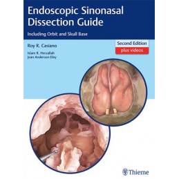
Superb multimedia resource provides clinical insights on endoscopic sinonasal dissection techniques
This remarkable manual encompasses the authors 30 years of experience and unique perspectives teaching endoscopic sinonasal surgery to residents and fellows. It also reflects a wealth of surgical pearls from rhinology and endoscopic skull base surgery experts on how to safely navigate through the nose, sinuses, orbit, and skull base.
Following a stepwise approach designed to mirror a residents progression in the cadaver lab, this user-friendly manual includes the most pertinent information on instrumentation, anteroposterior approaches, and postero-anterior approaches. Starting with the philosophy and history of sinus surgery, the reader is introduced to basic anatomical and surgical concepts - progressing to complete sphenoethmoidectomy and frontal sinusotomy. Subsequent chapters delineate advanced dissection techniques including dacryocystorhinostomy, orbital decompression, anterior skull base resection, infratemporal fossa approach, nasopharyngectomy, and skull base repair techniques utilizing grafts and local/regional flaps. Complementary external approaches to the frontal and maxillary sinuses are also illustrated.
Key Features
This visually-rich manual is ideal for residents in otolaryngology-head and neck surgery, as well as rhinology and endoscopic skull base fellows. It will also benefit otolaryngologists, ophthalmologists, and neurosurgeons who wish to brush up on specific endoscopic dissection techniques relative to their individual practice needs.
Data sheet
Section I Introduction to Endoscopic Sinonasal Surgical Landmarks
1. Introduction to Endoscopic Sinonasal Surgery
2. Anteroposterior versus Posteroanterior Approaches through the Paranasal Sinuses
3. The Use of Anatomical Landmarks: A Stepwise Approach to the Paranasal Sinuses
Section II Basic Endoscopic Sinonasal Surgical Anatomy and Techniques
4. Instrumentation and Operating Room Setup
5. Sinonasal and Skull Base CT Anatomy
6. Magnetic Resonance Sinonasal and Skull Base Anatomy
7. Endoscopic Intranasal Examination
8. Inferior Turbinoplasty and Submucous Resection of the Inferior Turbinate
9. Septoplasty
10. Middle Turbinoplasty
11. Uncinectomy and Middle Meatal Antrostomy
12. Anterior Ethmoidectomy
13. Posterior Ethmoidectomy
14. Sphenoid Sinusotomy
15. Retrograde Dissection Along the Skull Base for Advanced Sinonasal Disease
16. Frontal Sinusotomy
Section III Advanced Endoscopic Sinonasal, Orbital, and Skull Base Surgical Anatomy and Techniques
17. Olfactory Anatomy
18. Nasolacrimal System and Dacryocystorhinostomy
19. Orbital Decompression
20. Optic Nerve Decompression
21. Anterior and Posterior Ethmoid Arteries
22. Extended Maxillary Sinusotomies
23. Extended Frontal Sinusotomy and the Modified Lothrop Procedure
24. Extended Sphenoid Sinusotomy
25. Sphenopalatine Foramen, Pterygopalatine Fossa, and Vidian Canal
26. Transpterygoid Approaches to the Infratemporal Fossa and Meckels Cave
27. Approach to the Sella Turcica and Suprasellar Region
28. Lateral Sphenoid Sinus Wall, Internal Carotid Artery, and Adjacent Neurovascular Structures
29. Anterior Skull Base Resection
30. Approaches to the Clivus, Petrous Apex, Craniocervical Junction, and Odontoid Decompression
31. Approach to the Nasopharynx and the Parapharyngeal Space
32. Orbital Dissection
33. Nasoseptal and Inferior Turbinate Vascularized Flaps
34. Endoscopic Skull Base Reconstruction
35. Common Adjunctive External Skin Incisions for Approaches to the Frontal Maxillary Sinuses
36. External Approach to the Superior, Lateral, and Inferior Orbital Walls and Adjacent Paranasal Sinuses
37. Endoscopic Sinus Surgery Considerations in the Pediatric Population
