- Reduced price

Order to parcel locker

easy pay

 Delivery policy
Choose Paczkomat Inpost, Orlen Paczka, DHL, DPD or Poczta Polska. Click for more details
 Security policy
Pay with a quick bank transfer, payment card or cash on delivery. Click for more details
 Return policy
If you are a consumer, you can return the goods within 14 days. Click for more details
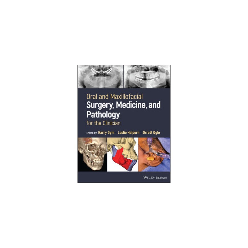
Single volume reference bringing together surgery, medicine, and pathology to provide relevant clinical information
Oral and Maxillofacial Surgery, Medicine, and Pathology for the Clinician presents key clinical information on oral surgery, medicine, and pathology in a single, easy-to-use resource, covering procedures performed in the dental clinic in a clear but concise manner and putting key details at the clinician’s fingertips.
Clinical scenarios are clearly described with treatment flow paths, and to enable seamless reader comprehension, charts and algorithms also support the text. The text focuses on essential office-related topics that are not esoteric but rather common in occurrence. The book speaks directly to topics of interest that will add value to the practitioner’s practice. Major surgical procedures not commonly performed by practicing oral surgeons are not included.
Overall, the text contains important up-to-date information that can be immediately put to use in clinical practice.
Oral and Maxillofacial Surgery, Medicine, and Pathology for the Clinician covers sample topics like::
The full scope of oral surgery is thoroughly covered in this multidisciplinary, current reference, making Oral and Maxillofacial Surgery, Medicine, and Pathology for the Clinician an essential tool for oral and maxillofacial surgeons, general dentists, and dental students looking to build upon their foundations of practical knowledge.
Data sheet
Contributors xvii
Preface xix
Part I Basics 1
1 Patient Evaluation and Management of Medical Problems in the Oral Surgery Patient 3
Risk Assessment 3
Documentation 4
Management of Patients with Medical Problems 5
Cardiovascular Disease 6
Hypertension 6
Angina Pectoris 7
Myocardial Infarction 8
Stroke/Cerebrovascular Accident 9
Cardiac Arrhythmias 9
Heart Failure 10
Treatment Guidelines 10
Endocrine Disorders 11
Diabetes Mellitus 11
Thyroid Disease 12
Adrenal Insufficiency 13
Hepatic Disease 13
Renal Disease 15
Dialyzed Patients 15
Pulmonary Disease 15
Asthma 15
Chronic Obstructive Pulmonary Disease 15
Pregnancy 16
Conclusion 18
References 18
2 Risk Reduction Strategies 21
Methods of Risk Reduction 21
Faulty Record Keeping 21
Informed Consent 21
Conclusion 23
Reference 23
3 Preparing the Dental Office for Medical Emergencies: Essentials of an Emergency Kit 25
Staff 25
Equipment 26
Oxygen 26
Airway Adjuncts 26
Automated External Defibrillators (Figure 3.2) 26
Vitals Monitoring 27
Intravenous Kits 27
Emergency Drug Kit 27
Oxygen 27
Aromatic Ammonia 27
Aspirin 28
Albuterol 28
Glucose 28
Nitroglycerin 28
Diphenhydramine 29
Epinephrine 29
Sedation-Specific Emergencies 29
Summary 29
References 29
Part II Dentoalveolar Surgery 31
4 Surgical Management of the Impacted Canine 33
Etiology 33
Diagnosis 33
Treatment and Management of the Impacted Canine 35
Goals 35
Interceptive Treatment to Prevent Impactions 35
Surgical Management of the Impacted Canine 35
Open vs Closed Surgery 35
Palatal Maxillary Impactions 36
Labial Maxillary Impactions 36
Mandibular Impactions 37
Complications 37
References 38
5 Crown Lengthening 39
Biologic Width 39
Indications for Crown Lengthening [3] 39
Contraindications for Crown Lengthening [1] 40
Procedures Carried Out Prior to Crown Lengthening [4] 40
Bone Sounding [2] 40
Sequence of Treatment for Crown Lengthening 40
External Bevel Gingivectomy 40
Internal Bevel Gingivectomy 40
Flap Surgery with Osseous Surgery 41
Apically Positioned Flap With or Without Osseous Surgery 42
Combined with Orthodontic Extrusion 42
Classification of Esthetic Crown Lengthening [2] 42
Postoperative Care [3] 43
References 43
Part III Implantology 45
6 Bone-Grafting Techniques and Biomaterials for Alveolar Ridge Augmentation 47
Bone Graft Materials and Healing Physiology 47
Introduction 47
Bone Graft and Tissue Engineering Materials – Outline 47
Autogenous (Natural) 47
Allograft (Natural) 47
Xenograft – Bovine, Porcine, Equine, Marine Coral, or Algal Sources 47
Alloplast 47
Other Synthetic Sources (Engineered) 48
Autologous Platelet Concentrate 48
Bone Graft and Tissue Engineering Materials 48
Autograft 48
Allograft 48
Mineralized Freeze-Dried Bone Allograft 49
Demineralized Freeze-Dried Bone Allograft 49
Particulate Cortical, Cancellous, and Corticocancellous Allograft 49
Xenograft 49
Natural Hydroxyapatite 49
Bio-Oss – Bovine Derived 49
Natural Hydroxyapatite – Marine Coral Derived 49
Calcium Carbonate – Biocoral 49
Fluorohydroxyapatite (FHA) – Natural Sea Algae Derived 50
Alloplast (Synthetic Sources) 50
Hydroxyapatite Based (Synthetic HA) 50
Marine Coral Derived (Hydroxyapatite) 50
Coralline Porous Hydroxyapatite – Interpore (Synthetic HA) 50
Coralline Porous Hydroxyapatite – Pro Osteon (Synthetic HA) 50
Nanocrystalline Hydroxyapatite (Synthetic HA) 51
Tricalcium Phosphate (Synthetic) 51
Biphasic HA and B-TCP Material (Synthetic Combination) 51
Calcium Sulfate – Gypsum Based (Synthetic) 51
Calcium Sulfate – Nanocrystalline (Synthetic) 51
Biphasic Calcium Sulfate (Synthetic) 51
Bioactive Glass Ceramics (Synthetic) 52
Other Synthetic Sources – Recombinant Bone Morphogenetic Protein (rhBMP) 52
Autologous Platelet Concentrates (See Table 6.1 for Complete Formulations) 52
Platelet-Rich Plasma 52
Platelet-Rich Fibrin 52
Ridge Preservation 54
Ridge Preservation Indications 54
Ridge Preservation Algorithm 54
Ridge Preservation Surgical Technique 55
Ridge Preservation Surgical Complications 57
Ridge Preservation Implant Survival and Success Rates 57
Ridge Preservation Conclusion (Box 6.4) 58
Guided Bone Regeneration (GBR) 59
Guided Bone Regeneration Indications 59
Nonabsorbable Membrane 59
Titanium Mesh 59
e-PTFE [51, 52] 59
Nonexpanded d-PTFE (Osteogenics Biomedical) [6, 49–51, 53] 59
Titanium-reinforced PTFE [51, 54] 60
Absorbable Membrane 61
Collagen Base (Bovine, Porcine, or Human Tendon, Dermis, Skin, or Pericardium) 61
Polymeric Membrane (Manufactured Synthetic Membrane) 61
Tuberosity Harvest Technique (Figures 6.9–6.11, Box 6.5) 62
Guided Bone Regeneration 62
Surgical Technique for Three Wall Defect (Figures 6.15–6.18) 62
Surgical Technique for Moderate-to-Severe Defect 62
Particulate Graft Resorption Post Grafting 64
Guided Bone Regeneration Complications 65
Guided Bone Regeneration Implant Survival and Success Rates 65
Guided Bone Regeneration Conclusion (Box 6.6) 65
Intraoral Onlay Graft 66
Indications (Particulate and Block Graft) 66
Subperiosteal Tunneling Technique with Absorbable Membrane 67
Autogenous Onlay Corticocancellous Grafts Healing 68
Surgical Harvest Technique: Intraoral Lateral Ramal Shelf and Symphysis (Figure 6.8) 68
Autogenous Intraoral Block Grafts – Surgical Complications 70
Allogeneic Block Bone (Cadaver Bone) 70
Block Graft Implant Survival and Success Rate 70
Block Graft Conclusion (Box 6.7) 70
Ridge Split 71
Indications 71
Ridge Split Surgical Technique 71
Ridge Split Complications 71
Ridge Split Implant Survival and Success Rates 72
Ridge Split Conclusion (Box 6.8) 72
Interpositional Bone Graft or “Sandwich Osteotomy” 73
Indications 73
Interpositional Bone Graft Surgical Technique 73
Interpositional Bone Graft Complications 74
Interpositional Bone Graft Implant Survival and Success Rates 74
Interpositional Bone Graft Conclusion (Box 6.9) 74
Distraction Osteogenesis 75
Indications 75
Distraction Osteogenesis Surgical Technique 77
Distraction Osteogenesis Complications 77
Distraction Osteogenesis Implant Survival and Success Rates 77
Distraction Osteogenesis Conclusion (Box 6.10) 77
Postoperative Instructions 77
Conclusion 78
Horizontal Augmentation Recap (Tables 6.5 and 6.6) 78
Horizontal Augmentation Complication Recap 78
Vertical Augmentation Recap (Tables 6.5 and 6.6) 79
References 81
7 Maxillary Sinus Augmentation 85
Introduction 85
Maxillary Sinus Anatomy 85
Indications, Contraindications, Limitations 85
Lateral Window Approach 86
Transalveolar (Crestal) Approach 86
Bone-Grafting Material 87
Complications 87
References 89
8 Technologic, Material, and Procedural Advancements in Dental Implant Surgery 91
Introduction 91
Three-Dimensional Imaging 91
Computerized Implant Planning Technology 92
Intraoral Optical Impressions and Integration with CBCT, CAD/CAM, and Stereolithography 92
Surgical Drilling Guide Integration and Fabrication 94
Guided Navigation in Osteotomy Preparation and Implant Placement 94
Membranes for Bone Grafting 95
BMP, PRGF, and PRP 96
Implant-Supported, Full-Arch, Fixed Prostheses with Immediate Loading and “All-on-Four” 97
Zygomatic Implants 98
Lasers 99
Conclusion 100
References 100
Part IV Trauma 101
9 Diagnosis and Management of Dentoalveolar Trauma 103
Introduction 103
Evaluation 103
History 103
Physical Examination 103
Radiographic Studies 104
Diagnosis and Management of Dentoalveolar Injuries 104
Injuries to the Dental Hard Tissue and Pulp 104
Crown Infraction 104
Crown Fracture 104
Crown-Root Fracture 105
Root Fracture 105
Injuries to the Periodontal Tissues 105
Concussion 105
Subluxation 105
Intrusion 105
Extrusion 106
Lateral Luxation 106
Avulsion 106
Dentoalveolar Injuries in the Primary Dentition 107
Splinting 108
Injuries to the Gingiva or Oral Mucosa 108
Injuries to Supporting Bone 108
Follow-Up 109
Conclusion 109
References 109
Part V Pathology 113
10 Biopsy Technique: When, Where, and How? 115
Introduction 115
Patient Evaluation: Health History, Medications 115
Lesion History 116
Clinical Examination 117
Indications for Biopsies 120
Precancerous Lesion: “Potentially Malignant Disorders” 120
Biopsy Techniques 123
Incisional Biopsy 123
Excisional Biopsy 124
Punch Biopsy 124
Adjunctive Techniques 124
Lugol’s Iodine 126
Toluidine Blue 126
Brush Biopsy/Cytology 128
References 128
11 Diagnosis and Management of Recurrent Lesions of the Oral Mucosa 131
Introduction 131
Aphthous Lesions and Recurrent Aphthous Stomatitis 131
Presentation 131
Etiology 131
Diagnosis 132
Treatment 132
Herpetic Lesions 133
Course of the Disease 133
Diagnosis 133
Treatment 133
Candidiasis 134
Clinical Presentation 134
Diagnosis 134
Treatment 134
Lichen Planus 135
Clinical Presentation 135
Diagnosis 135
Treatment 135
Pemphigus Vulgaris 136
Clinical Presentation 136
Etiology 136
Diagnosis 137
Treatment 137
Erythema Multiforme 137
Clinical Presentation 137
Diagnosis 138
Treatment 138
Fixed Drug Eruptions 138
Clinical Presentation 138
Etiology 138
Diagnosis 139
Treatment 139
References 139
12 Benign Pediatric Pathology: Diagnosis and Management 143
Introduction 143
Eruption Cyst 144
Ameloblastoma 145
Aneurysmal Bone Cyst 146
Melanotic Neuroectodermal Tumor of Infancy 147
Juvenile Ossifying Fibroma 148
Cherubism 149
Hemangioma 150
Vascular Malformation 151
Verrucous Vulgaris and Condyloma Acuminatum 152
References 153
13 Diagnosis and Management of Salivary Gland Pathology 157
Introduction 157
Management of Sialolithiasis 158
Bacterial Salivary Gland Infections 159
Viral Sialadenitis 160
Granulomatous Disease of the Salivary Gland 160
Autoimmune Diseases 161
Warthin Tumor 162
Conclusion 163
References 163
14 Odontogenic Cysts and Odontogenic Tumors 167
Introduction 167
Developmental Odontogenic Cysts 168
Odontogenic Keratocyst (Keratocystic Odontogenic Tumor) 169
Lateral Periodontal Cyst and Botryoid Odontogenic Cyst 171
Gingival Cyst 172
Orthokeratinized Odontongenic Cyst 173
Ameloblastoma 174
Clinical Presentation 175
Histologic Features 176
Squamous Odontogenic Tumor 177
Histologic Features 178
Histologic Features 179
References 180
15 Osteomyelitis of the Jaw 183
Classification 183
Laboratory Analysis 184
Microbiology 186
Treatment 187
Case Presentations 188
Osteomyelitis of the Maxilla 188
Osteomyelitis of the Mandible 189
References 192
16 Obstructive Sleep Apnea 193
Introduction 193
Symptoms 194
Surgical Procedures 195
Uvulopalatopharyngoplasty 196
Genial Tubercle Advancement 197
Conclusion 198
References 198
17 Temporomandibular Disorders: A Clinician’s Guide for Nonsurgical and Surgical Interventions 201
Introduction 201
Nonsurgical Treatment Approaches 204
Methods of Therapy 205
Maxillary and Mandibular Full-Arch Splints 206
Surgical Approaches 207
Case Presentation 214
Conclusion 216
References 216
18 Postoperative Complications in Oral Surgery 219
Complications Arising During the Procedure 219
Root Fracture and Displacement 220
Nerve Injury 221
Complications after Oral Surgery 222
Infection 223
Pain, Swelling, and Trismus 224
References 225
19 Odontogenic Infections: Anatomy, Etiology, and Treatment 227
Introduction 227
Physical Examination 228
Radiographic Imaging 229
Pain Control 230
Treatment Techniques 231
Method 231
Palatal Abscess 232
Method 233
Buccal and Midfacial Abscess 234
Method 235
Methods 236
Pterygomandibular Space 237
Pericoronitis 238
Antibiotic Therapy 239
References 240
Part VI Pain Control 243
20 Approaches to the Management of Facial Pain 245
Introduction 245
Diagnostic Approach to Facial Pain Patients 246
Neuropathic Pain 248
Episodic Neuropathic Pain 249
Continuous NP 250
Neurovascular Pain 253
Topical NSAIDs 254
Injection Therapy 255
Complications of Injection Therapy 256
Summary and Future Directions 257
References 257
21 Local Anesthesia: Agents and Techniques 261
Local Anesthetics 261
Nerve Block Techniques 262
Greater Palatine Approach 263
Sigmoid Notch Approach 264
Halstead Method 265
Akinosi Technique (Figure 21.6) 266
Auxilliary/Supplemental Techniques 267
Intrasulcular Infusion 268
Trigger Point Injections 270
Nasal Spray 271
References 272
22 Nitrous Oxide 275
History 275
Use and Administration 275
Contraindications 276
Safety Mechanisms 277
References 278
Part VII Oral Medicine 281
23 Antibiotic Prophylaxis in Oral and Maxillofacial Surgery: Recent Trends in Therapeutic Applications 283
Introduction 283
Infective Endocarditis 285
Additional Considerations about Infective Endocarditis Antibiotic Prophylaxis (When Indicated) 286
Third Molars/Extractions 287
Cleft lip and palate 289
Dental Implants 290
Trauma 291
Conclusion 292
References 292
24 Management of Patients on Anticoagulation 295
Introduction 295
Heparins 296
Antiplatelet Agents 297
Conclusion 299
References 299
25 Burning Mouth Syndrome 301
Introduction 301
Etiology and Risk Factors 302
Diagnosis 303
Treatment 304
Primary Burning Mouth Syndrome 305
Conclusion 306
References 307
Index 311
Reference: 81412
Author: John Breeze
Reference: 13036
Author: Jatin P. Shah
komplet
Reference: 93929
Author: Dania F Tamimi