- Reduced price

Order to parcel locker

easy pay

 Delivery policy
Choose Paczkomat Inpost, Orlen Paczka, DHL, DPD or Poczta Polska. Click for more details
 Security policy
Pay with a quick bank transfer, payment card or cash on delivery. Click for more details
 Return policy
If you are a consumer, you can return the goods within 14 days. Click for more details
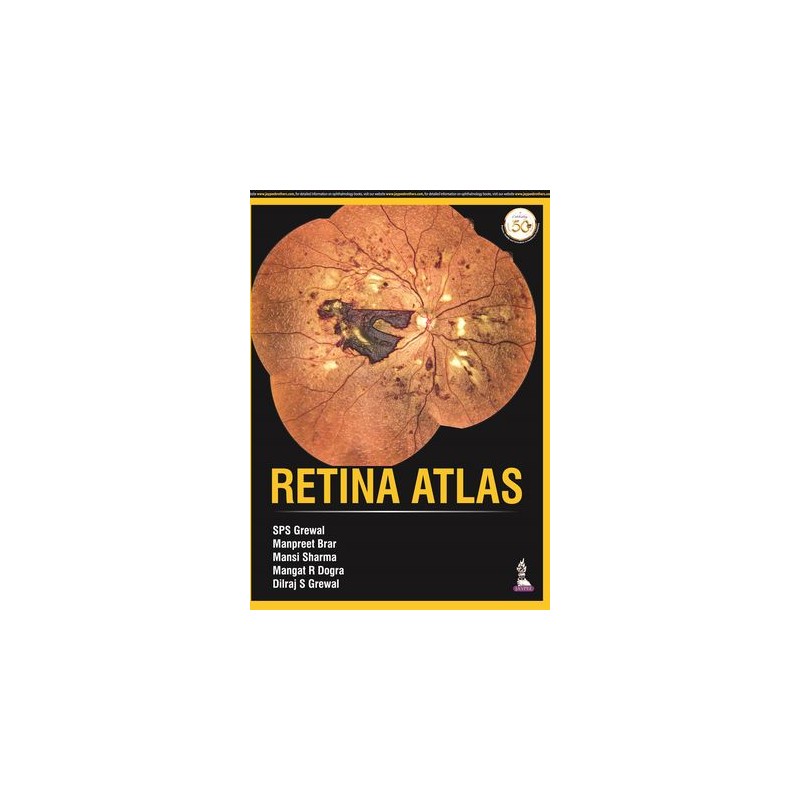
This atlas provides ophthalmologists with a collection of images to help with the identification, diagnosis and subsequent treatment of retinal disorders.
The images are procured from Eidon scanner technology and also include optical coherence tomography (OCT) pictures to assist with understanding of related pathologies.
Divided into nine sections, the book begins with images illustrating the normal fundus. Each of the following sections covers a different retinal disorder including diabetic retinopathy, macula disorders, retinal detachment, ocular tumours and hereditary diseases.
Each section features a multitude of images, each with brief descriptive text to assist understanding.
Key points
Data sheet
1. Normal Fundus
2. Diabetic Retinopathy
3. Retinal Vascular Disorders
4. Macula
5. Retinal Detachment
6. Inflammatory
7. Hereditary
8. Ocular Tumours and Optic Nerve Disorders
9. Miscellaneous
Reference: 82448
Author: Jeffrey A. Nerad
A Personal Tutorial
Reference: 93175
Author: David B. Samimi