- Reduced price

Order to parcel locker

easy pay

 Delivery policy
Choose Paczkomat Inpost, Orlen Paczka, DHL, DPD or Poczta Polska. Click for more details
 Security policy
Pay with a quick bank transfer, payment card or cash on delivery. Click for more details
 Return policy
If you are a consumer, you can return the goods within 14 days. Click for more details
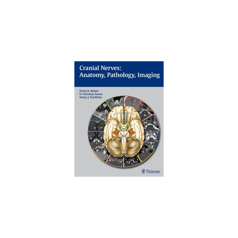
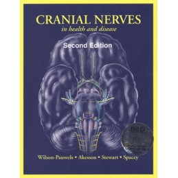
A single-volume resource for detailed coverage of the anatomy, function, and pathology of the cranial nerves with CT and MRI correlation
This beautifully illustrated book combines a detailed exposition of the anatomy and function of the cranial nerves with practical coverage of clinical concepts for the assessment and differential diagnosis of cranial nerve dysfunction. An introductory chapter provides a brief overview of cranial nerve anatomy and function, skull base anatomy, classification of pathologies, and imaging approaches. Each of the twelve chapters that follow is devoted to in-depth coverage of a different cranial nerve. These chapters open with detailed discussion of the various functions of each nerve and normal anatomy. The authors then describe common lesions and present a series of cases that are complemented by CT images and MRIs to illustrate disease entities that result in cranial nerve dysfunction.
Features
This book is an indispensable reference for practicing physicians and trainees in neurosurgery, neurology, neuroradiology, radiology, and otolaryngology-head and neck surgery. It will also serve as a valuable resource for students seeking to gain a solid understanding of the anatomy, function, and pathology of the cranial nerves.
Data sheet
Reference: 54564
Author: Dennis Marchiori
With Skeletal, Chest, & Abdominal Pattern Differentials
Reference: 102705
Author: Sanjay Mallya
Principles and Interpretation
Reference: 93660
Author: Lorrie L. Kelley