- Reduced price

Order to parcel locker

easy pay

 Delivery policy
Choose Paczkomat Inpost, Orlen Paczka, DHL, DPD or Poczta Polska. Click for more details
 Security policy
Pay with a quick bank transfer, payment card or cash on delivery. Click for more details
 Return policy
If you are a consumer, you can return the goods within 14 days. Click for more details
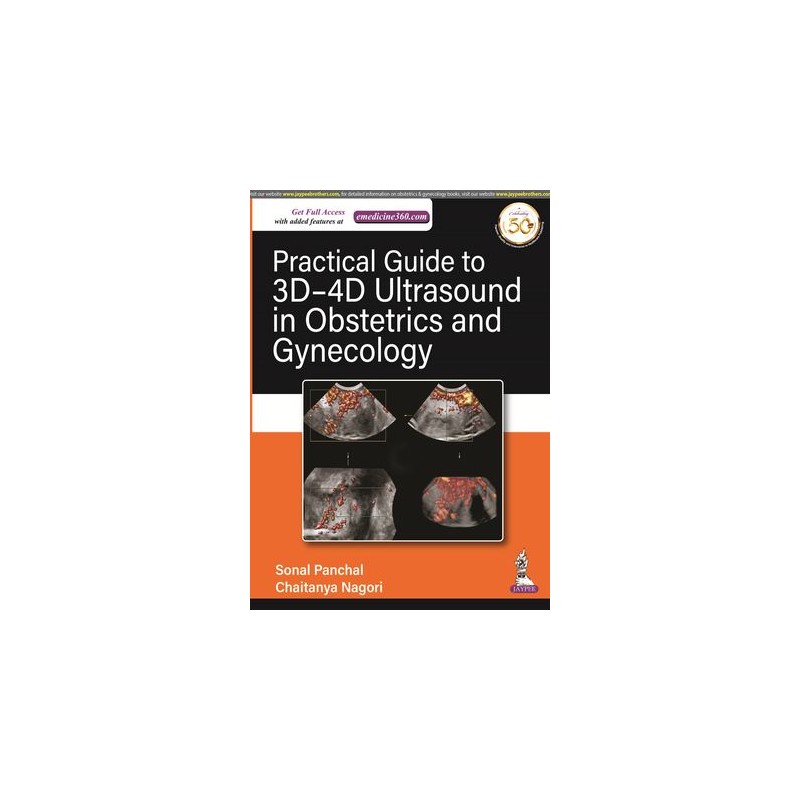
3D ultrasound shows a still image of a foetus, far more detailed than the 2D flat grey scale imaging. 4D ultrasound is more advanced, showing a moving image, allowing obstetricians to evaluate foetal well-being. It is also used by gynaecologists to examine uterine anomalies.
This book is a practical guide to the use of 3D and 4D ultrasound in obstetrics and gynaecology.
Divided into three sections, the text begins with an introduction to ultrasound, its working and application, its function, software, and volume calculation tools.
Section Two covers clinical applications of volume ultrasound in obstetrics, explaining its use during the first trimester, for foetal abnormalities, for functional assessment of foetal brain development, and in labour.
The final section discusses the application of ultrasound in gynaecology, covering uterine abnormalities, adnexal lesions, and in infertility.
The book concludes with an appendix detailing different terms used by different brands.
Key points
Data sheet
Section 1:: 3D Ultrasound Basics
Section 2:: Clinical Applications of Volume Ultrasound in Obstetrics
Section 3:: Clinical Applications of Volume Ultrasound in Gynecology and Infertility