- Reduced price

Order to parcel locker

easy pay


 Delivery policy
Choose Paczkomat Inpost, Orlen Paczka, DHL, DPD or Poczta Polska. Click for more details
 Security policy
Pay with a quick bank transfer, payment card or cash on delivery. Click for more details
 Return policy
If you are a consumer, you can return the goods within 14 days. Click for more details
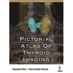
Pictorial Atlas of Thyroid Imaging is a concise, highly illustrated guide from an Abu Dhabi based editorial team of radiology and endocrinology experts.
Divided into eight chapters, the book introduces the basics of interpretation of thyroid and parathyroid ultrasound images, as well as providing more advanced guidance for practitioners with experience of these techniques. The first chapter covers the thyroid gland, including normal thyroid and post thyroidectomy changes, followed by a range of conditions which might require imaging, such as congenital disorders, nodular diseases, thyroiditis and hyperthyroidism.
Each chapter covers different lesions of the thyroid gland, with accompanying images and in-depth descriptions to assist fundamental radiology reporting skills. The book concludes with a chapter on the parathyroid glands and a miscellaneous chapter.
Enhanced by 750 high quality ultrasound and pathology images, and images of tumour types, Pictorial Atlas of Thyroid Imaging is an excellent resource for interns, endocrinologists and endocrine surgeons.
Key Points
Data sheet
1. The Thyroid Gland
1.1 Normal Thyroid
1.2 Post Thyroidectomy Changes
2. Congenital Disorders of the Thyroid
3. Goiter
4. Nodular Diseases of the Thyroid
4.1 Benign Looking Nodules
4.2 Suspicious Nodules
4.3 Lymph Nodes
4.4 Thyroid Cancer Residual/Recurrent Disease
5. Thyroiditis
6. Hyperthyroidism
7. Parathyroid Glands
8. Miscellaneous
Index
Reference: 6259
Author: John Emory Campbell
