- Reduced price

Order to parcel locker

easy pay


 Delivery policy
Choose Paczkomat Inpost, Orlen Paczka, DHL, DPD or Poczta Polska. Click for more details
 Security policy
Pay with a quick bank transfer, payment card or cash on delivery. Click for more details
 Return policy
If you are a consumer, you can return the goods within 14 days. Click for more details
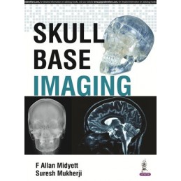
This book is a complete guide to skull base imaging covering all current techniques including CT, MRI, Ultrasound, Angiography, CT Cisternography and Plain Film Radiography.
Divided into eight sections, each part covers imaging techniques for a different region of the skull base, including cerebellopontine, anterior, posterior and middle cranial fossa, the pituitary region, craniovertebral junction and more. The final chapters discuss inflammatory disorders and sarcomas.
The book includes the very latest techniques including diffusion weighted imaging (DWI) and FLAIR. Each chapter features key points, clues, incidence and location, as well as detail on presentation, epidemiology, pathology, staging, differential diagnosis, treatment and prognosis.
Authored by recognised experts from Washington and Michigan, this comprehensive guide is enhanced by radiological images and illustrations.
Key Points
Data sheet
Section I:: Pituitary Region
Section II:: Cerebellopontine Angle
Section III:: Anterior Cranial Fossa
Section IV:: Middle Cranial Fossa
Section V:: Craniovertebral Junction
Section VI:: Posterior Cranial Fossa
Section VII:: Inflammatory
Section VIII:: Sarcomas
