- Reduced price

Order to parcel locker

easy pay


 Delivery policy
Choose Paczkomat Inpost, Orlen Paczka, DHL, DPD or Poczta Polska. Click for more details
 Security policy
Pay with a quick bank transfer, payment card or cash on delivery. Click for more details
 Return policy
If you are a consumer, you can return the goods within 14 days. Click for more details
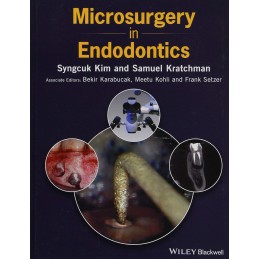
Microsurgery in Endodontics provides the definitive reference to endodontic microsurgery, with instructive photographs and illustrations.
Data sheet
Contributors xiii
Preface xv
Acknowledgements xvii
1 The Dental Operating Microscope 1
Frank Setzer
1.1 Benefits of the Operating Microscope 1
1.2 Key Features of Operating Microscopes 1
1.3 Customizing a Microscope 3
1.3.1 Light Source 3
1.3.2 Documentation 5
1.3.3 Individual Microscope Adjustment (Parfocaling) 5
Suggested Readings 7
2 Microsurgical Instruments 9
SeungHo Baek and Syngcuk Kim
2.1 Examination Instruments 9
2.2 Incision and Elevation Instrument 9
2.3 Tissue Retraction Instruments 11
2.4 Osteotomy Instruments 14
2.5 Curettage Instruments 14
2.6 Inspection Instruments 15
2.7 Ultrasonic Units and Tips for Root End Preparation 17
2.8 Microplugger Instruments 20
2.9 Suturing Instruments 20
2.10 Miscellaneous Instruments 22
3 Medication-Related Osteonecrosis of the Jaw and Endodontic Microsurgery 25
Chafic Safi and Bekir Karabucak
Suggested Readings 29
4 Indications and Contraindications 31
Bekir Karabucak and Garrett Guess
4.1 Introduction 31
4.2 Surgical Success Dependent on Ability to Perform Ideal Protocols 31
4.3 Etiology Assessment through Examination and Treatment 31
4.4 Periodontal Considerations and Surgery 33
4.5 Influential Patient Factors 33
4.6 Condition of Previous Endodontic Treatment 37
Suggested Readings 38
5 Anesthesia and Hemostasis 39
Siva Rethnam-Haug, Aleksander Iofin, and Syngcuk Kim
5.1 Armamentarium 39
5.1.1 Epinephrine 39
5.2 Presurgical Phase 40
5.2.1 Administration of Local Anesthetic 40
5.2.2 Injection Techniques 40
5.2.3 Topical Anesthesia 41
5.2.4 Additional Techniques 41
5.2.5 Maxillary Anesthesia 41
5.2.6 Mandibular Anesthesia 43
5.2.7 Bilateral Mandibular Surgery 43
5.3 Surgical Phase 44
5.3.1 Topical Hemostatic Agents 44
5.3.1.1 Epinephrine Pellets 45
5.3.1.2 Ferric Sulfate 45
5.4 Summary of Hemostatic Techniques in Endodontic Microsurgery 46
5.5 Postsurgical Phase 46
Suggested Readings 48
6 Flap Design in Endodontic Microsurgery 49
Francesco Maggiore and Frank Setzer
6.1 Armamentarium 49
6.1.1 Flap Outline 49
6.1.2 Papilla Management 52
6.1.3 Incision 54
6.1.4 Flap Elevation 54
6.1.5 Flap Retraction 55
Suggested Readings 56
7 Osteotomy 57
Francesco Maggiore and Syngcuk Kim
7.1 Armamentarium 57
7.2 Osteotomy 57
7.2.1 Distinction between Bone and Root Tip 57
7.2.2 Clinical Situations for Endodontic Microsurgery 58
7.3 Intact Cortical Plate without a Radiographic Periapical Lesion 59
7.4 Intact Cortical Plate with a Periapical Lesion 60
7.5 Fenestration through the Cortical Plate Leading to the Apex 60
7.5.1 Optimal Osteotomy Size 61
7.5.2 Key Hole Osteotomy Modification 61
7.5.3 BoneWindow Technique 61
8 Root End Resection 67
Spyros Floratos, Fouad Al-Malki, and Syngcuk Kim
8.1 Armamentarium 67
8.2 Root End Resection 67
8.3 Root End Resection: Steep Bevel versus Shallow Bevel 69
Suggested Readings 72
9 Inspection of the Resected Root Surface: Importance of Isthmus 73
Spyros Floratos, Jorge Vera, Fouad Al-Malki, and Syngcuk Kim
9.1 Armamentarium 73
9.1.1 Methylene Blue Staining (MBS) 73
9.1.2 Isthmus 77
9.1.3 Types of Isthmus 77
9.1.4 Incidence 77
9.1.5 Histological Findings of Isthmus 79
9.1.6 Clinical Significance and Management 81
Suggested Readings 82
10 Ultrasonic Root End Preparation 83
Spyros Floratos and Syngcuk Kim
10.1 Armamentarium 83
Suggested Readings 89
11 MTA and Bioceramic Root End Filling Materials 91
Sujung Shin, Ian Chen, Bekir Karabucak, SeungHo Baek, and Syngcuk Kim
11.1 Mineral Trioxide Aggregate (MTA) 92
11.1.1 Advantages of MTA 92
11.1.1.1 Sealing Ability 92
11.1.1.2 Biocompatibility and Bioactivity 92
11.1.2 Drawbacks of MTA 93
11.2 Bioceramics 93
11.3 MTA and Bioceramic Application During Apicoectomy 95
11.4 Other Types of Cements for Root End Filling 96
11.4.1 Intermediate Restorative Material (IRM) 97
11.4.2 Super Ethoxybenzoic Acid (SuperEBA) 97
11.4.3 Geristore and Retroplast 98
11.4.4 New Types of Cements for Root End Filling 98
Suggested Readings 98
12 Flap Reposition and Suturing 101
Francesco Maggiore and Meetu Kohli
12.1 Suture Removal 101
Suggested Readings 111
13 PeriapicalWound Healing 113
Ingrida Dapkute, Georges Bandelac, Chafic Safi, and Frank Setzer
13.1 Principles ofWound Healing 113
13.2 Healing after Apical Microsurgery 113
13.3 Incomplete Healing/Scar Formation 113
13.4 Evaluation of Healing after Apical Surgery 114
13.5 Healing Evaluation Using CBCT 115
Suggested Readings 118
14 Cone Beam Computed Tomography 119
Garrett Guess, Fouad Al-Malki,Meetu Kohli, Bekir Karabucak, and Samuel Kratchman
14.1 How CBCT Works 120
14.2 Indications and Clinical Applications 120
Suggested Readings 142
15 Mental Nerve Management 143
Paula Mendez-Montalvo, Fouad Al-Malki, and Syngcuk Kim
15.1 Armamentarium 143
15.1.1 Mental Foramen and Nerve 143
15.1.1.1 Location 143
15.1.1.2 Anterior Loop 143
15.1.1.3 Number of Mental Foramina 144
15.1.2 Mental Foramen Detection on Radiographs 144
15.1.2.1 Periapical Radiograph 144
15.1.2.2 Panoramic Films 145
15.1.2.3 Cone Beam Computed Tomography (CBCT) 145
15.1.3 Neurosensory Alteration 145
15.1.3.1 Surgical Technique to Avoid Iatrogenic Mental Nerve Trauma and Injury 146
15.1.4 Groove Technique Using Piezoelectric Surgery 146
Suggested Readings 150
16 Maxillary Posterior Surgery, the Sinus, andManaging Palatal Access 151
Garrett Guess and Samuel Kratchman
16.1 Maxillary Premolars 151
16.1.1 Access 151
16.1.2 Instrumentation 151
16.2 Sinus Exposure 151
16.3 Maxillary First Molars 153
16.3.1 Access 153
16.3.2 Palatal Approach 155
16.4 Second Molars 159
16.4.1 Periodontal Aspects 161
Suggested Readings 162
17 Surgical Root Perforation Repair 163
Raed Kasem, Samuel Kratchman, and Meetu Kohli
17.1 Possible Challenges to Non-surgical Perforation Repair 163
17.2 Factors that Enhance Positive Long-Term Prognosis for Perforation Repair 164
17.3 Surgical Perforation Repair Techniques 166
17.4 Surgical Treatment for External Root Resorption 168
Suggested Readings 177
18 Intentional Replantation 179
David Li and Samuel Kratchman
18.1 Armamentarium 179
18.1.1 Success Rate 179
18.1.2 Indications 179
18.1.3 Replantation or Apicoectomy 179
18.1.4 Extraction 182
18.1.5 Extraoral Phase 183
18.1.6 Storage Medium 183
18.1.7 Replantation 183
18.1.8 Splinting 185
18.1.9 Postop Instructions 186
18.1.10 Cone Beam CT Scan 186
18.1.11 Repairing Procedural Mishaps 186
Suggested Readings 191
19 Guided Tissue Regeneration in Endodontic Microsurgery 193
Garrett Guess and Samuel Kratchman
Suggested Readings 202
20 Implants versus Endodontic Microsurgery 205
Frank Setzer and Syngcuk Kim
20.1 Historical Perspective 205
20.2 Benefits of Implants 205
20.3 Long-Term Prognosis of Dental Implants 205
20.4 Implant Complications 206
20.5 Long-Term Prognosis of Endodontically Treated Teeth with Root End Surgery 207
20.6 Conclusion 209
Suggested Readings 210
21 Prognosis of Endodontic Microsurgery 213
Meetu Kohli and Euiseong Kim
21.1 Best Available Evidence 213
21.2 Parameters for Success: Clinical and Radiographic 2D 214
21.3 Parameters for Success: “Penn 3D Criteria” for Assessing Healing on CBCT 215
21.4 Reversal of Success 215
21.5 TraditionalMethods 217
21.6 Modern Technique versus the Complete Microsurgical Approach 218
21.7 Root End Filling Materials 218
21.8 Case Selection 219
21.9 Resurgery 219
21.10 Summary 219
Suggested Readings 219
22 Positioning 221
Samuel Kratchman and Syngcuk Kim
22.1 Armamentarium 221
Suggested Readings 226
Index 227
