- Reduced price

Order to parcel locker

easy pay


 Delivery policy
Choose Paczkomat Inpost, Orlen Paczka, DHL, DPD or Poczta Polska. Click for more details
 Security policy
Pay with a quick bank transfer, payment card or cash on delivery. Click for more details
 Return policy
If you are a consumer, you can return the goods within 14 days. Click for more details
Fluorescein angiography is an eye test that uses a special dye and camera to look at blood flow in the retina and choroid, the two layers in the back of the eye (MedlinePlus).
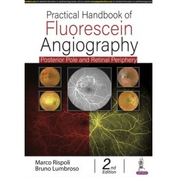
This book is a practical guide to the interpretation of fluorescein angiography images and subsequent diagnosis of retinal disorders.
The second edition has been fully revised and updated to provide the latest advances in the field. New topics including retinal periphery and ocular oncology, and new images have been added.
Divided into seven sections, the book covers interpretation of normal and pathological fluorescein angiography, new, dyeless imaging for retina and choroid (OCT and OCT Angiography), pathological fluorescein angiography analytical study, pathological fluorescein angiography synthesis, major fluorescein angiography syndromes posterior pole, study of retinal periphery and the future of fluorescein angiography.
Written by internationally recognised specialists Marco Rispoli and Bruno Lumbroso from Rome Eye Hospital and Centro Oftalmologico Mediterraneo, this handbook is highly illustrated with angiograph images and tables to help trainees and clinicians recognise and interpret angiographic findings and make an accurate diagnosis of ophthalmic disorders.
The previous edition (9789350909911) published in 2014.
Data sheet
PART I:: INTERPRETING A NORMAL AND PATHOLOGICAL FLUORESCEIN ANGIOGRAPHY
Chapter 1 Retinal anatomy and fluorescein angiography
Chapter 2 Normal fluorescein angiography of the central retina
Chapter 3 Normal fluorescein angiogram of the optic disk
Chapter 4 Normal fluorescein angiography of the choroid
Chapter 5 Interpreting a pathological fluorescein angiography
PART II:: NEW DYELESS IMAGING FOR RETINA AND CHOROID STUDY:: OCT AND OCT ANGIOGRAPHY
Chapter 6 Assessment of fluorescein and indocyanine angiography versus OCT-angiography::
Fluorescein angiography/OCT-angiography:: A progressive transition
PART III:: PATHOLOGICAL FLUORESCEIN ANGIOGRAPHY ANALYTICAL STUDY
Chapter 7 Abnormal hyperfluorescence
Chapter 8 Abnormal hypofluorescence
Chapter 9 Abnormalities in circulation time
PART IV:: PATHOLOGICAL FLUORESCEIN ANGIOGRAPHY-SYNTHESIS
Chapter 10 Synthetic evaluation
PART V:: MAJOR FLUORESCEIN ANGIOGRAPHY SYNDROMES POSTERIOR POLE
Chapter 11 Diabetic retinopathy
Chapter 12 Age-related macular degeneration and other macular degenerations
Chapter 13 Vascular occlusions
Chapter 14 Pachychoroid spectrum of disorders:: Retinal epitheliopathies
Chapter 15 Vitreoretinal interface syndrome
Chapter 16 Diagnosis of a dark area in the fluorescein angiography
Chapter 17 Inflammatory disorders
PART VI:: FLUORESCEIN ANGIOGRAPHY STUDY OF RETINAL PERIPHERY
Chapter 18 Fluorescein angiography of the retinal periphery
Chapter 19 Retinal periphery in diabetic retinopathy
Chapter 20 Widefield angiography in ocular oncology
PART VII:: CONCLUSION
Chapter 21 The future of fluorescein angiography
Index
Reference: 55706
Author: Timo Krings
Reference: 9166
Author: Stephen Rollnick
Jak pomóc pacjentom w zmianie złych nawyków i ryzykownych zachowań
Reference: 50220
Author: Anita Agarwal
2-Volume Set - Expert Consult: Online and Print
Reference: 95785
Author: Suber S Huang
