- Reduced price

Order to parcel locker

easy pay


 Delivery policy
Choose Paczkomat Inpost, Orlen Paczka, DHL, DPD or Poczta Polska. Click for more details
 Security policy
Pay with a quick bank transfer, payment card or cash on delivery. Click for more details
 Return policy
If you are a consumer, you can return the goods within 14 days. Click for more details
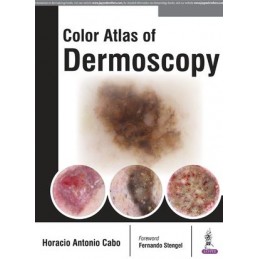
Dermoscopy is a non-invasive, widely used diagnostic tool that aids the diagnosis of skin lesions and is proven to increase the accuracy of melanoma diagnosis.
This colour atlas is a comprehensive guide to the diagnosis of skin lesions and melanomas using a dermoscope.
Beginning with an introduction to the use of the dermascope, the following chapters teach clinicians how to recognise dermoscopic criteria, colours and patterns, how to diagnose different types of lesions and calculate diagnostic algorithms.
The finals sections cover related topics including entomodermatoscopy, inflammatoscopy, trichoscopy and capilaroscopy.
This highly useful resource is enhanced by more than 1000 clinical images and illustrations.
Key points
Data sheet
Reference: 36913
Author: Joseph Varon
Year Book Handbooks Series
