- Reduced price

Order to parcel locker

easy pay

 Delivery policy
Choose Paczkomat Inpost, Orlen Paczka, DHL, DPD or Poczta Polska. Click for more details
 Security policy
Pay with a quick bank transfer, payment card or cash on delivery. Click for more details
 Return policy
If you are a consumer, you can return the goods within 14 days. Click for more details
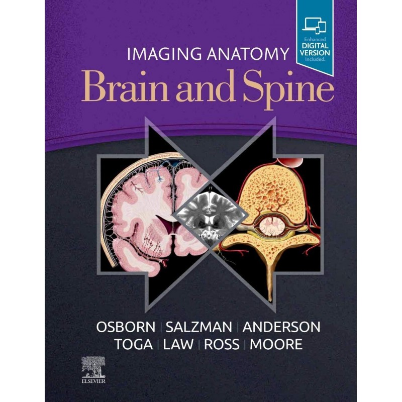
This richly illustrated and superbly organized text/atlas is an excellent point-of-care resource for practitioners at all levels of experience and training. Written by global leaders in the field, Imaging Anatomy:: Brain and Spine provides a thorough understanding of the detailed normal anatomy that underlies contemporary imaging. This must-have reference employs a templated, highly formatted design; concise, bulleted text; and state-of- the-art images throughout that identify the clinical entities in each anatomic area.
Data sheet
Brain
Scalp, Skull, and Meninges
Scalp and Calvarial Vault
Cranial Meninges
Pia and Perivascular Spaces
Supratentorial Brain Anatomy
Cerebral Hemispheres Overview
Gyral/Sulcal Anatomy
White Matter Tracts
Basal Ganglia and Thalamus
Other Deep Gray Nuclei
Limbic System
Sella, Pituitary, and Cavernous Sinus
Pineal Region
Primary Somatosensory Cortex (Areas 1, 2, 3)
Primary Motor Cortex (Area 4)
Superior Parietal Cortex (Areas 5, 7)
Premotor Cortex and Supplementary Motor Area (Area 6)
Superior Prefrontal Cortex (Area 8)
Dorsolateral Prefrontal Cortex (Areas 9, 46)
Frontal Pole (Area 10)
Orbitofrontal Cortex (Area 11)
Insula and Parainsula Areas (Areas 13, 43)
Primary Visual and Visual Association Cortex (Areas 17, 18, 19)
Temporal Cortex (Areas 20, 21, 22)
Posterior Cingulate Cortex (Areas 23, 31)
Anterior Cingulate Cortex (Areas 24, 32, 33)
Subgenual Cingulate Cortex (Area 25)
Retrosplenial Cingulate Cortex (Areas 29, 30)
Parahippocampal Gyrus (Areas 28, 34, 35, 36)
Fusiform Gyrus (Area 37)
Temporal Pole (Area 38)
Inferior Parietal Lobule (Areas 39, 40)
Primary Auditory and Auditory Association Cortex (Areas 41, 42)
Inferior Frontal Gyrus (Areas 44, 45, 47)
High-Resolution Cortical Anatomy
Brain Network Anatomy
Functional Network Overview
Neurotransmitter Systems
Default Mode Network
Attention Control Network
Sensorimotor Network
Visual Network
Limbic Network
Language Network
Memory Network
Social Network
Infratentorial Brain
Brainstem and Cerebellum Overview
Midbrain
Pons
Medulla
Cerebellum
Cerebellopontine Angle/IAC
CSF Spaces
Ventricles and Choroid Plexus
Subarachnoid Spaces/Cisterns
Skull Base and Cranial Nerves
Skull Base Overview
Anterior Skull Base
Central Skull Base
Posterior Skull Base
Cranial Nerves Overview
Olfactory Nerve (CNI)
Optic Nerve (CNII)
Oculomotor Nerve (CNIII)
Trochlear Nerve (CNIV)
Trigeminal Nerve (CNV)
Abducens Nerve (CNVI)
Facial Nerve (CNVII)
Vestibulocochlear Nerve (CNVIII)
Glossopharyngeal Nerve (CNIX)
Vagus Nerve (CNX)
Accessory Nerve (CNXI)
Hypoglossal Nerve (CNXII)
Extracranial Arteries
Aortic Arch and Great Vessels
Cervical Carotid Arteries
Intracranial Arteries
Intracranial Arteries Overview
Intracranial Internal Carotid Artery
Circle of Willis
Anterior Cerebral Artery
Middle Cerebral Artery
Posterior Cerebral Artery
Vertebrobasilar System
Veins and Venous Sinuses
Intracranial Venous System Overview
Dural Sinuses
Superficial Cerebral Veins
Deep Cerebral Veins
Posterior Fossa Veins
Extracranial Veins
Spine
Vertebral Column, Discs, and Paraspinal Muscle
Vertebral Column Overview
Ossification
Vertebral Body and Ligaments
Intervertebral Disc and Facet Joints
Paraspinal Muscles
Craniocervical Junction
Cervical Spine
Thoracic Spine
Lumbar Spine
Sacrum and Coccyx
Cord, Meninges, and Spaces
Spinal Cord and Cauda Equina
Meninges and Compartments
Vascular
Spinal Arterial Supply
Spinal Veins and Venous Plexus
Plexi and Peripheral Nerves
Brachial Plexus
Lumbar Plexus
Sacral Plexus and Sciatic Nerve
Peripheral Nerve and Plexus Overview
Reference: 48591
Author: Peter M. Som
Expert Consult- Online and Print