- Obniżka

Medycyna estetyczna

Diagnoza i psychoterapia uzależnień

Zamawiaj do paczkomatu

Płać wygodnie

 Dostawa
Wybierz Paczkomat Inpost, Orlen Paczkę, DHL, DPD, Pocztę, email (dla ebooków). Kliknij po więcej
 Płatność
Zapłać szybkim przelewem, kartą płatniczą lub za pobraniem. Kliknij po więcej szczegółów
 Zwroty
Jeżeli jesteś konsumentem możesz zwrócić towar w ciągu 14 dni*. Kliknij po więcej szczegółów
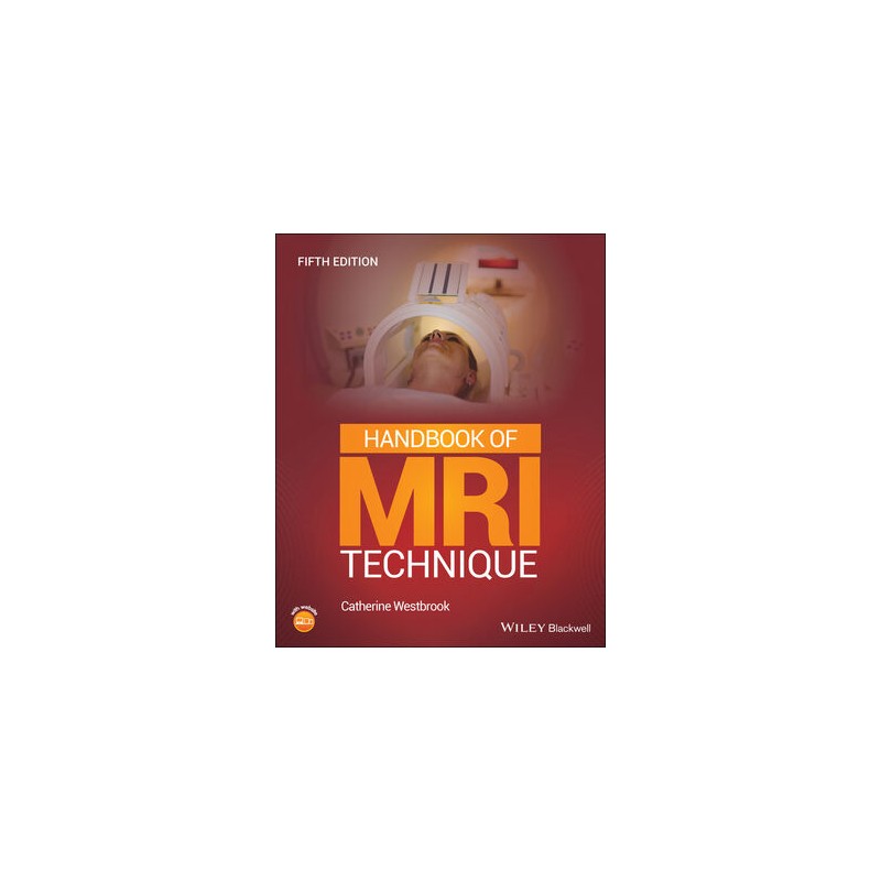
Distinguished educator Catherine Westbrook delivers a comprehensive and intuitive resource for radiologic technologists in this newly revised Fifth Edition of the Handbook of MRI Technique. With a heavy emphasis on protocol optimisation and patient care, the book guides the uninitiated through scanning techniques and assists more experienced technologists with image quality improvement.
The new edition includes up-to-date scanning techniques and an additional chapter on paediatric imaging. The latest regulations on MRI safety are referenced and there are expanded sections on slice prescription criteria. The book also includes the contributions of several clinical experts, walking readers through key theoretical concepts, discussing practical tips on cardiac gating, equipment use, patient care, MRI safety, and contrast media. Step-by-step instruction is provided on scanning each anatomical area, complete with patient positioning and image quality optimisation techniques.
The book includes::
Perfect for radiography students and newly qualified practitioners, as well as practitioners preparing for MRI-based certification and examination, the Handbook of MRI Technique will also prove to be an invaluable addition to the libraries of students in biomedical engineering technology and radiology residents.
Opis
Contributors ix
Preface xi
Acknowledgements xiii
About the Companion Website xv
Chapter 1 How to Use This Book 1
Introduction 1
Common indications 2
Basic anatomy 2
Equipment 2
Patient positioning 5
Slice prescription 6
Suggested protocol 6
Protocol optimization 6
Patient considerations 6
Contrast usage 6
Summary 7
Terms and abbreviations used in Part 2 7
Conclusion 17
Part 1 Theoretical and Practical Concepts 19
Chapter 2 Protocol Parameters and Trade-offs 21
Introduction 21
Signal-to-noise ratio (SNR) 24
Contrast-to-noise ratio (CNR) 24
Spatial resolution 25
Scan time 26
Decision strategies 27
Conclusion 28
Chapter 3 Pulse Sequences 29
Introduction 29
Conventional spin echo (CSE) 29
Fast spin echo or turbo spin echo (FSE/TSE) 32
Inversion recovery (IR/FSE/TSE-IR) 34
Rewound gradient echo 35
Balanced GRE 35
Spoiled GRE 36
Reverse echo GRE 37
Echo planar imaging (EPI) 38
Conclusion 42
Chapter 4 Flow Phenomena and Artefacts 44
Flow phenomena 44
Introduction 44
Time of flight (TOF) 45
Entry slice phenomenon 46
Intra-voxel dephasing 46
Flow artefact remedies 46
Artefacts 48
Introduction 48
Phase mismapping 48
Aliasing 48
Chemical shift 49
Out-of-phase signal cancellation 49
Truncation 49
Magnetic susceptibility 49
Magic angle 50
Conclusion 51
Chapter 5 Gating and Respiratory Compensation Techniques 53
Introduction 53
Cardiac gating (ECG/EKG gating) 53
Peripheral gating (Pe gating) 58
Ciné imaging 59
Imaging planes 60
Respiratory Compensation (RC) 60
Conclusion 61
Chapter 6 Patient Care and Safety 62
Introduction 62
Patient screening 62
Safety zones 63
Safety concerns during the examination 64
Patient counselling 65
Patient immobilization 67
Patient after-care 67
Conclusion 67
Chapter 7 Contrast Agents 68
Introduction 68
Gd-based positive contrast agents 68
Negative contrast agents 70
Patient considerations 70
Conclusion 70
Part 2 Examination Areas 71
Chapter 8 Head and Neck 73
Brain 73
Temporal lobes 86
Internal auditory meatus and posterior fossa 92
Pituitary fossa 97
Orbits 102
Paranasal sinuses 109
Pharynx 113
Larynx 119
Thyroid and parathyroid glands 124
Salivary glands 129
Temporomandibular joints 133
Vascular imaging 137
Head and neck imaging – key points 141
Chapter 9 Spine 142
Cervical spine 142
Thoracic spine 151
Lumbar spine 156
Whole spine imaging 162
Spine imaging – key points 166
Chapter 10 Chest 167
Lungs and mediastinum 167
Heart and great vessels 175
Thymus 186
Breast 189
Axilla 199
Brachial plexus 202
Chest imaging – key points 206
Chapter 11 Abdomen 207
Liver and biliary system 207
Kidneys and adrenal glands 215
Pancreas 222
Bowel 227
Vascular imaging 232
Abdominal imaging – key points 235
Chapter 12 Pelvis 236
Prostate 236
Rectum and testes 244
Uterus and cervix 247
Pelvic imaging – key points 251
Chapter 13 Upper Limb 252
Shoulder 252
Humerus 262
Elbow 268
Forearm 276
Wrist and hand 281
Upper limb imaging – key points 288
Chapter 14 Lower Limb 289
Hips 289
Femur 298
Knee 303
Tibia and fibula 311
Ankle 316
Foot 323
Vascular imaging 328
Lower limb imaging – key points 334
Chapter 15 Paediatric Imaging 335
Introduction 335
Creating the right environment 335
Sedation and anaesthesia 337
The MRI examination 345
Conclusion 375
Paediatric imaging – key points 375
Index 376