- Obniżka

Medycyna estetyczna

Diagnoza i psychoterapia uzależnień

Zamawiaj do paczkomatu

Płać wygodnie

 Dostawa
Wybierz Paczkomat Inpost, Orlen Paczkę, DHL, DPD, Pocztę, email (dla ebooków). Kliknij po więcej
 Płatność
Zapłać szybkim przelewem, kartą płatniczą lub za pobraniem. Kliknij po więcej szczegółów
 Zwroty
Jeżeli jesteś konsumentem możesz zwrócić towar w ciągu 14 dni*. Kliknij po więcej szczegółów
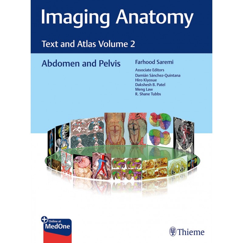
Unique anatomic atlas provides an indispensable virtual desk dissection experience
Normal imaging anatomy and variants, including diagnostic and surgical anatomy, are the cornerstones of radiologic knowledge. Imaging Anatomy:: Text and Atlas Volume 2, Abdomen and Pelvis is the second in a series of four richly illustrated radiologic references edited by distinguished radiologist Farhood Saremi. The atlas is coedited by esteemed colleagues Damian Sanchez-Quintana, Hiro Kiyosue, Dakshesh B. Patel, Meng Law, and R. Shane Tubbs with contributions from an impressive cadre of international authors. Succinctly written text and superb images provide readers with a virtual, user-friendly dissection experience.
This exquisitely crafted atlas combines fundamental core anatomy principles with modern imaging and postprocessing methods to increase understanding of intricate anatomical features. Twenty-two concise chapters cover the abdominal wall, alimentary tract, liver, biliary system, pancreas, spleen, peritoneum, genitourinary system, pelvic floor, neurovasculature, and surface anatomy. Relevant anatomical components of the abdomen and pelvis are discussed, including musculature, arteries, veins, lymphatics, ducts, and innervation.
Key Highlights
This unique resource provides an excellent desk reference for differentiating normal versus pathologic anatomy. It is essential reading for medical students, radiology residents and veteran radiologists, internists, and general surgeons, as well as vascular and transplant surgeons.
This print book includes complimentary access to a digital copy on https://medone.thieme.com.
Publishers Note:: Products purchased from Third Party sellers are not guaranteed by the publisher for quality, authenticity, or access to any online entitlements included with the product.
Opis
1. Abdominopelvic Wall
2. Esophagus
3. Stomach
4. Small Intestine
5. Colon
6. Liver
7. Spleen
8. Biliary System
9. Pancreas
10. Mesenteric Vasculature of Lower Gastrointestinal System
11. Portal Venous System
12. Peritoneal Spaces
13. Adrenal Glands, Kidneys, Ureters, and Bladder
14. Extraperitoneal Space
15. Male Genitourinary
16. Female Genital System
17. Perineum
18. Pelvic Floor
19. Abdominal Aorta and Major Branches
20. Systemic Veins of the Abdomen and Pelvis
21. Lymphatics of the Abdomen, Pelvis, and Perineum
22. Surface Anatomy and Projectional Surface Anatomy
Indeks: 90374
Autor: Farhood Saremi
Indeks: 100868
Autor: Meng Law
Indeks: 104746
Autor:
Indeks: 75822
Autor: Hanns-Georg Klein