- Obniżka

Medycyna estetyczna

Diagnoza i psychoterapia uzależnień

Zamawiaj do paczkomatu

Płać wygodnie

 Dostawa
Wybierz Paczkomat Inpost, Orlen Paczkę, DHL, DPD, Pocztę, email (dla ebooków). Kliknij po więcej
 Płatność
Zapłać szybkim przelewem, kartą płatniczą lub za pobraniem. Kliknij po więcej szczegółów
 Zwroty
Jeżeli jesteś konsumentem możesz zwrócić towar w ciągu 14 dni*. Kliknij po więcej szczegółów
BMA Book Awards - Winner of Basic and Clinical Sciences category!
The perfect up-to-date imaging guide for a complete and 3-dimensional understanding of applied human anatomy
Imaging is ever more integral to anatomy education and throughout modern medicine. Building on the success of previous editions, this fully revised sixth edition provides a superb foundation for understanding applied human anatomy, offering a complete view of the structures and relationships within the whole body, using the very latest imaging techniques.
All relevant imaging modalities are included, from plain radiographs to more advanced imaging of ultrasound, CT, MRI, functional imaging and angiography. Coverage is further enhanced by a carefully selected range of BONUS electronic content, including clinical photos and cases, ultrasound videos, labelled radiograph slidelines, cross-sectional imaging stacks and test-yourself materials. Uniquely, key syllabus image sets are now highlighted throughout to aid efficient study, as well as the most common, clinically important anatomical variants that you should be aware of.
This superb package is ideally suited to the needs of medical students, as well as radiologists, radiographers and surgeons in training. It will also prove invaluable to the range of other students and professionals who require a clear, accurate, view of anatomy in current practice.
Labelled image stacks-that allow you to review cross-sectional imaging as if using an imaging workstation
Labelled image slidelines-showing features in a full range of body radiographs to enhance understanding of anatomy in this essential modality
Self-test image slideshows with multi-tier labelling-to aid learning and cater for beginner to more advanced experience levels
Labelled ultrasound videos-bring images to life, reflecting this increasingly clinically practiced technique
Questions and answers accompany each chapter-to test your understanding and aid exam preparation
34 pathology tutorials-based around nine key concepts and illustrated with hundreds of additional pathology images, to further develop your memory of anatomical structures and lead you through the essential relationships between normal and abnormal anatomy High-yield USMLE topics-clinical photos and cases for key topics, linked and highlighted in chapters
Opis
Indeks: 102778
Autor: Eric A. Storch
Indeks: 13590
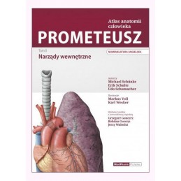
Autor: Michael Schuenke
angielska nomenklatura
Indeks: 17401
Autor: red. wyd. pol. Bogdan Ciszek
angielska i polska nomenklatura