- Obniżka

Medycyna estetyczna

Diagnoza i psychoterapia uzależnień

Zamawiaj do paczkomatu

Płać wygodnie

 Dostawa
Wybierz Paczkomat Inpost, Orlen Paczkę, DHL, DPD, Pocztę, email (dla ebooków). Kliknij po więcej
 Płatność
Zapłać szybkim przelewem, kartą płatniczą lub za pobraniem. Kliknij po więcej szczegółów
 Zwroty
Jeżeli jesteś konsumentem możesz zwrócić towar w ciągu 14 dni*. Kliknij po więcej szczegółów
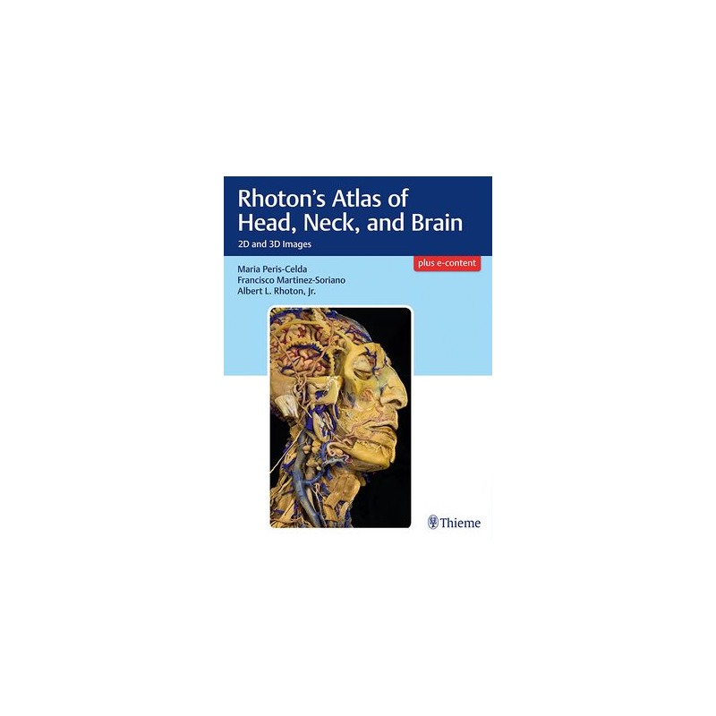
Masterful 2D and 3D head, neck, and brain dissections provide unsurpassed insights into head, neck, and brain anatomy
An internationally renowned and beloved author, educator, brain anatomist, and neurosurgeon, Professor Albert Rhoton has a special place in medical history. He was revered by students and colleagues and is regarded as one of the fathers of modern microscopic neurosurgery. A driving principle in his anatomy lab was the simple phrase, Every Second. This was embraced in his philosophy that every second of every day, a patients life was improved by a surgeon assisted by the anatomic knowledge his lab helped elucidate and distribute.
Rhotons Atlas of Head, Neck, and Brain is the visually exquisite crowning achievement of Dr. Rhotons brilliant career and unwavering dedication to the intertwined pursuits of surgical anatomy and neurosurgery. The atlas reflects the unparalleled contributions Dr. Rhoton made to the contemporary understanding of neurosurgical anatomy. Dr. Peris-Celda, with the collaboration of an impressive cadre of international multidisciplinary experts, worked closely under Dr. Rhotons tutelage on this project. This book is the culmination of 5 years of work and experience gleaned from more than 40 years of surgical anatomy research and exquisite dissection techniques performed in Dr. Rhotons laboratory.
Special Features
Breathtakingly stunning, this atlas is certain to be a treasured reference for medical students, residents, and clinicians specializing in neurosurgery, facial plastic surgery, otolaryngology, maxillofacial surgery, and craniofacial surgery for many years to come.
Opis
Part I Osteology of the Head and Neck
1 Adult and Fetal Skull
2 Bones of the Skull and Skull Bone Articulations
3 Cervical Vertebrae
Part II Face and Neck
4 Face: Superficial Dissection
5 Face: Deep Dissection
6 Anterior Aspect of the Neck: Deep Dissection
7 Anterior Aspect of the Neck: Deep Dissection
8 Posterior Aspect of the Neck
9 Parapharyngeal Dissection
Part III Ear, Nose, Pharynx, Larynx, and Orbit
10 External and Middle Ear
11 Internal Ear
12 Nose: Sagittal Dissection
13 Nose: Coronal Dissection
14 Endonasal Endoscopy
15 Pharynx
16 Larynx
17 Orbit
18 Eye and Orbital Contents
19 Internal Structure of the Eyeball
Part IV Neuroanatomy and Cranial Base
20 Cerebrum
21 Cerebellum and Brainstem
22 Brain, Meninges, and Sutures
23 Cerebrovascular and Intraventricular Dissection
24 Cranial Base and Craniocervical Junction
25 Sagittal and Endoscopic Dissection of the Cranial Base
26 Cranial Nerves
27 Brain Sections
28 Fiber Dissection of the Brain
Indeks: 100962
Autor: Elizabeth Parker