• Zamawiaj do paczkomatu
  • Płać wygodnie
  • Obniżka
  • Nowy
Seven Cavernomas: Tenets and Techniques for Resection

Seven Cavernomas: Tenets and Techniques for Resection

9781684204946
856,80 zł
771,12 zł Zniżka 85,68 zł Brutto
Najniższa cena w okresie 30 dni przed promocją: 771,12 zł
Ilość
Zapowiedź - cena orientacyjna
2025-12-17

  Dostawa

Wybierz Paczkomat Inpost, Orlen Paczkę, DHL, DPD, Pocztę, email (dla ebooków). Kliknij po więcej

  Płatność

Zapłać szybkim przelewem, kartą płatniczą lub za pobraniem. Kliknij po więcej szczegółów

  Zwroty

Jeżeli jesteś konsumentem możesz zwrócić towar w ciągu 14 dni*. Kliknij po więcej szczegółów

Opis

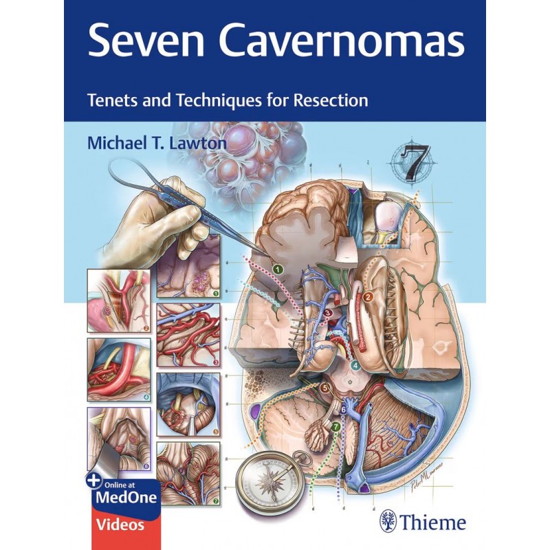
An incomparable collection of evidence-based cavernoma road trips through the cerebral terrain


Seven Cavernomas:: Tenets and Techniques for Resection is the fourth volume in a remarkable series by internationally renowned neurosurgeon Michael T. Lawton. As with the three prior volumes, Dr. Lawton leverages his vast expertise as a leading cerebrovascular neurosurgeon, sharing insights and knowledge gleaned from operating on over 1,400 cavernomas. Seven Cavernomas integrates quintessential clinical, anatomical, and microsurgical concepts into a comprehensive heuristic to position neurosurgeons to achieve the best patient outcomes with cavernoma microsurgical resection.


Section I details the 10 tenets of cavernoma resection, starting with a taxonomy for classification by location and surface presentation. Subsequent chapters describe brainstem and cerebral anatomy as well as dissection techniques in detail, covering the triangle concept, arterial landmarks, hotspots of brain eloquence, recurrence, patient selection, and neurosurgical cartography. Section II examines the seven types of cavernous malformations, with insightful pearls of wisdom for resection techniques. The closing chapter concludes with a discussion of the future role neurosurgery will play in understanding how the brain gives us our consciousness, emotion, memory, and intelligence.


Key Highlights


• Taxonomy of 7 cavernoma types and 35 subtypes guides the neurosurgeon to choose the optimal approaches, execute the operation skillfully, and maximize intraoperative performance


• Brainstem cavernous malformation cartography maps the special relationships among craniotomy, approach, anatomical triangles, safe entry zones, and arteries as vascular waypoints


• An impressive compendium of 65 surgical videos and 8 animations captures the action, progression, movement, and technical nuances that sculpt the art of neurosurgery


• A total of 700+ exquisitely rendered illustrations and clinical images delineate anatomical


components with stunning accuracy


This volume is an essential reference for every vascular neurosurgeon. The book demonstrates in meticulous detail how the art and science of map-making is a path to crystallizing the art and science of cavernoma resection. The taxonomy provides a consistent nomenclature for discussion while providing technical and navigational nuance, inspiring confidence, and empowering neurosurgeons to improve patient outcomes.

Szczegóły produktu
Georg Thieme
104136
9781684204946
9781684204946

Opis

Rok wydania
2025
Numer wydania
1
Oprawa
twarda
Liczba stron
452
Wymiary (mm)
215.90 x 279.40

    1. Section I The Tenets
    2. 1 Taxonomy
    3. 1.1 Classifying Surgical Pathology
    4. 1.2 Informing Surgical Strategy
    5. 1.3 Neuroanatomy
    6. 1.4 Neuroradiology
    7. 1.5 Neurology
    8. 1.6 Neurosurgery
    9. 1.7 Seven Cavernomas Framework
    10. 2 Subarachnoid Dissection

      1. 2.1 Seven Surgical Corridors
      2. 2.1.1 Cerebral Sulci
      3. 2.1.2 Sylvian Fissure
      4. 2.1.3 Interhemispheric Fissure
      5. 2.1.4 Ventricular System
      6. 2.1.5 Tentorial Fissure
      7. 2.1.6 Cerebellopontine Cistern
      8. 2.1.7 Cisterna Magna

    11. 2.2 Conclusion
    12. 3 Triangle Concept
    13. 3.1 The Triangle Concept
    14. 3.2 A System of Anatomical Triangles
    15. 3.3 Midbrain Triangles

      1. 3.3.1 Carotid-Oculomotor Triangle
      2. 3.3.2 Oculomotor-Tentorial Triangle
      3. 3.3.3 Supracerebellar-Supratrochlear and Supracerebellar-Infratrochlear Triangles
      4. 3.3.4 Infragalenic Triangle

    16. 3.4 Pontine Triangles

      1. 3.4.1 Posteromedial (Kawase) Triangle
      2. 3.4.2 Glossopharyngeal-Cochlear Triangle
      3. 3.4.3 Supra- and Infratrigeminal Triangles
      4. 3.4.4 Interlobular Triangle

    17. 3.5 Medullary Triangles

      1. 3.5.1 Vertebrobasilar Junctional Triangle
      2. 3.5.2 Subtonsillar Triangle
      3. 3.5.3 Vagoaccessory Triangle
      4. 3.5.4 Cerebellar Vallecular Triangle

    18. 3.6 Thalamic and Basal Ganglial Triangles

      1. 3.6.1 Supracarotid-Infrafrontal Triangle
      2. 3.6.2 Caudate-Thalamostriate Triangle
      3. 3.6.3 Septocaudate Triangle
      4. 3.6.4 Infragalenic Triangle

    19. 3.7 Cerebral and Cerebellar Triangles

      1. 3.7.1 Vallecular Triangle
      2. 3.7.2 Interlobular Triangle

    20. 3.8 Clinical Evidence
    21. 3.9 Conclusion
    22. 4 Arterial Landmarks
    23. 4.1 Rivers of the Brain
    24. 4.2 Middle Cerebral Arteries
    25. 4.3 Anterior Cerebral Arteries
    26. 4.4 Internal Carotid Artery
    27. 4.5 Posterior Cerebral Arteries
    28. 4.6 Superior Cerebellar Artery
    29. 4.7 Anterior Inferior Cerebellar Artery
    30. 4.8 Posterior Inferior Cerebellar Artery
    31. 4.9 Arterial Dissection Codes
    32. 5 Safe Entry Zones
    33. 5.1 From Inoperable to Operable
    34. 5.2 Clinical Evidence
    35. 5.3 The Safety of Safe Entry Zones
    36. 5.4 The 21 Brainstem Safe Entry Zones
    37. 5.5 Midbrain Safe Entry Zones

      1. 5.5.1 Interpeduncular Zone
      2. 5.5.2 Anterior Mesencephalic Zone
      3. 5.5.3 Lateral Mesencephalic Sulcus Zone
      4. 5.5.4 Intercollicular Zone
      5. 5.5.5 Supracollicular and Infracollicular Zones

    38. 5.6 Pontine Safe Entry Zones

      1. 5.6.1 Supratrigeminal and Infratrigeminal
      2. 5.6.2 Middle Cerebellar Peduncle or Lateral Pontine
      3. 5.6.3 Area Acustica
      4. 5.6.4 Median Sulcus (Pons)
      5. 5.6.5 Suprafacial Collicular
      6. 5.6.6 Superior Foveal
      7. 5.6.7 Pontomedullary Sulcus

    39. 5.7 Medullary Safe Entry Zones

      1. 5.7.1 Anterolateral Sulcus
      2. 5.7.2 Olive
      3. 5.7.3 Posterolateral Sulcus and Lateral Medullary
      4. 5.7.4 Median Sulcus (Medulla)
      5. 5.7.5 Infrafacial Collicular
      6. 5.7.6 Posterior Median Sulcus
      7. 5.7.7 Posterior Intermediate Sulcus

    40. 5.8 Accuracy of Preoperative MRI in Determining Surface Proximity
    41. 5.9 Neuronavigation, Hemosiderin Stain, and Neuromonitoring
    42. 5.10 Brainstem CM Taxonomy and Associated SEZs
    43. 6 Resection Technique
    44. 6.1 The Mulberry
    45. 6.2 Extracapsular Resection Technique
    46. 6.3 Intracapsular Resection Technique
    47. 6.4 Brain Transgression
    48. 6.5 Technical Overview of the Trans-MCP Approach
    49. 6.6 Superomedial Trajectory Beyond the SEZ: Superior Cerebellar Peduncle
    50. 6.7 Posteromedial Trajectory Beyond the SEZ: Pontine Tegmentum
    51. 6.8 Invisible Triangles
    52. 6.9 Orienteering Beyond
    53. 7 Eloquent Noneloquence
    54. 7.1 Ten Percent Myth
    55. 7.2 Eloquent Noneloquence
    56. 7.3 Clinical Evidence
    57. 7.4 Eloquent Cortex
    58. 7.5 Large-Scale Brain Networks
    59. 7.6 Seven Hotspots of Cerebral Eloquence
    60. 7.7 Expanding the Concept of Cerebral Eloquence
    61. 8 Residual and Recurrent Cavernous Malformations
    62. 8.1 Problem of Recurrence
    63. 8.2 Clinical Evidence
    64. 8.3 Detection
    65. 8.4 Surgical Blind Spots
    66. 8.5 Right-Angle Method
    67. 8.6 Fine Line
    68. 9 Patient Selection
    69. 9.1 Rationale for a Brainstem Cavernous Malformation Grading Scale
    70. 9.2 Lawton Brainstem CM Grading Scale
    71. 9.3 Elements of the Brainstem CM Grading System

      1. 9.3.1 Size
      2. 9.3.2 Crossing the Axial Midpoint
      3. 9.3.3 Developmental Venous Anomaly
      4. 9.3.4 Age
      5. 9.3.5 Hemorrhage

    72. 9.4 Validation of the Brainstem CM Grading System
    73. 9.5 Clinical Application
    74. 9.6 Giant Cavernomas
    75. 9.7 Hannegan’s Sign
    76. 10 Neurosurgical Cartography
    77. 10.1 Jackson Hole
    78. 10.2 Cartography
    79. 10.3 Maps as Metaphors for the Seven Cavernomas
    80. 10.4 Seven Cavernomas Maps
    81. 10.5 Maps for Safe Passage
    82. 10.6 Maps for Education
    83. 10.7 Maps for Exploration
    84. 10.8 The Metaphor
    85. Section II The Seven Cavernomas
    86. 11 Superficial Cerebral Cavernous Malformations
    87. 11.1 Introduction
    88. 11.2 Neuroanatomy of Superficial Cerebral Cavernous Malformation Subtypes

      1. 11.2.1 Convexity
      2. 11.2.2 Medial
      3. 11.2.3 Basal
      4. 11.2.4 Sylvian

    89. 11.3 Clinical Evidence
    90. 11.4 Resection Strategies for Superficial Cerebral Cavernous Malformations

      1. 11.4.1 Convexity Subtype
      2. 11.4.2 Medial Subtype
      3. 11.4.3 Basal Subtype
      4. 11.4.4 Sylvian Subtype

    91. 11.5 Superficial Cerebral Cavernous Malformations and Approach Selection
    92. 11.6 Approach Technique
    93. 11.7 Presenting Symptom
    94. 11.8 Clinical Syndromes
    95. 11.9 Conclusion
    96. 12 Basal Ganglia Cavernous Malformations
    97. 12.1 Introduction
    98. 12.2 Neuroanatomy of Three Basal Ganglia Cavernous Malformation Subtypes

      1. 12.2.1 Caudate
      2. 12.2.2 Putaminal
      3. 12.2.3 Pallidal

    99. 12.3 Clinical Evidence
    100. 12.4 Basal Ganglia Cavernous Malformation Subtypes

      1. 12.4.1 Caudate
      2. 12.4.2 Putaminal
      3. 12.4.3 Pallidal

    101. 12.5 Conclusion
    102. 13 Thalamic Cavernous Malformations
    103. 13.1 Introduction
    104. 13.2 Neuroanatomy of Thalamic Cavernous Malformation Subtypes

      1. 13.2.1 Anterior
      2. 13.2.2 Medial
      3. 13.2.3 Lateral
      4. 13.2.4 Choroidal
      5. 13.2.5 Pulvinar
      6. 13.2.6 Geniculate

    105. 13.3 Clinical Evidence
    106. 13.4 Resection Strategies for Thalamic Cavernous Malformation Subtypes

      1. 13.4.1 Anterior
      2. 13.4.2 Medial
      3. 13.4.3 Lateral
      4. 13.4.4 Choroidal
      5. 13.4.5 Pulvinar
      6. 13.4.6 Geniculate

    107. 13.5 Iterative Advances
    108. 13.6 Conclusion
    109. 14 Midbrain Cavernous Malformations
    110. 14.1 Introduction
    111. 14.2 Neuroanatomy of the Five Midbrain Cavernous Malformation Subtypes

      1. 14.2.1 Interpeduncular
      2. 14.2.2 Peduncular
      3. 14.2.3 Tegmental
      4. 14.2.4 Quadrigeminal
      5. 14.2.5 Periaqueductal

    112. 14.3 Clinical Evidence
    113. 14.4 Resection Strategies for Midbrain Cavernous Malformations

      1. 14.4.1 Interpeduncular
      2. 14.4.2 Peduncular
      3. 14.4.3 Tegmental
      4. 14.4.4 Quadrigeminal
      5. 14.4.5 Periaqueductal

    114. 14.5 Conclusion
    115. 15 Pontine Cavernous Malformations
    116. 15.1 Introduction
    117. 15.2 Neuroanatomy of the Six Pontine Cavernous Malformation Subtypes

      1. 15.2.1 Basilar
      2. 15.2.2 Peritrigeminal
      3. 15.2.3 Middle Peduncular
      4. 15.2.4 Inferior Peduncular
      5. 15.2.5 Rhomboid
      6. 15.2.6 Supraolivary

    118. 15.3 Clinical Evidence
    119. 15.4 Resection Strategies for Pontine Cavernous Malformations

      1. 15.4.1 Basilar Subtype
      2. 15.4.2 Peritrigeminal Subtype
      3. 15.4.3 Middle Peduncular Subtype
      4. 15.4.4 Inferior Peduncular Subtype
      5. 15.4.5 Rhomboid Subtype
      6. 15.4.6 Supraolivary Subtype

    120. 15.5 Signs and Syndromes
    121. 16 Medullary Cavernous Malformations
    122. 16.1 Introduction
    123. 16.2 Neuroanatomy of the Five Medullary Cavernous Malformation Subtypes

      1. 16.2.1 Pyramidal
      2. 16.2.2 Olivary
      3. 16.2.3 Cuneate
      4. 16.2.4 Gracile
      5. 16.2.5 Trigonal

    124. 16.3 Clinical Evidence
    125. 16.4 Resection Strategies for Medullary Cavernous Malformations

      1. 16.4.1 Pyramidal Subtype
      2. 16.4.2 Olivary Subtype
      3. 16.4.3 Cuneate Subtype
      4. 16.4.4 Gracile Subtype
      5. 16.4.5 Trigonal Subtype

    126. 16.5 Conclusion
    127. 17 Cerebellar Cavernous Malformations
    128. 17.1 Introduction
    129. 17.2 Neuroanatomy of the Six Cerebellar Cavernous Malformation Subtypes

      1. 17.2.1 Suboccipital
      2. 17.2.2 Tentorial
      3. 17.2.3 Petrosal
      4. 17.2.4 Vermian
      5. 17.2.5 Tonsillar
      6. 17.2.6 Deep Nuclear

    130. 17.3 Clinical Evidence
    131. 17.4 Resection Strategies for Cerebellar Cavernous Malformations

      1. 17.4.1 Suboccipital, Vermian, and Tonsillar
      2. 17.4.2 Tentorial
      3. 17.4.3 Petrosal
      4. 17.4.4 Deep Nuclear

    132. 17.5 Conclusion
    133. 18 Seven Cavernomas: Project Connectomunculus and the Mind
    134. 18.1 Seven Cavernomas Cartography
    135. 18.2 The Connectomunculus
    136. 18.3 The Mind
    137. 18.4 Gallery of Functional Networks
    138. 19 Suggested Readings
    139. Contributors
    140. Index
Zobacz także
Komentarze (0)