- Obniżka

Medycyna estetyczna

Diagnoza i psychoterapia uzależnień

Zamawiaj do paczkomatu

Płać wygodnie

 Dostawa
Wybierz Paczkomat Inpost, Orlen Paczkę, DHL, DPD, Pocztę, email (dla ebooków). Kliknij po więcej
 Płatność
Zapłać szybkim przelewem, kartą płatniczą lub za pobraniem. Kliknij po więcej szczegółów
 Zwroty
Jeżeli jesteś konsumentem możesz zwrócić towar w ciągu 14 dni*. Kliknij po więcej szczegółów
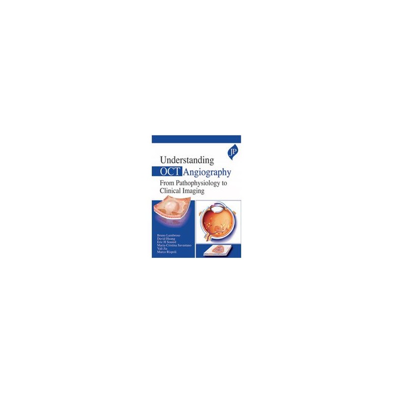
This handbook provides ophthalmologists and trainees with the latest advances in the rapidly developing field of OCT Angiography.
Beginning with an overview of the technology and terminology, the next chapter explains how to perform an OCTA examination. The following sections describe the pathophysiology, clinical features, imaging and management techniques for different retinal disorders.
Authored by an internationally recognised group of experts, led by Professor Bruno Lumbroso, the book is highly illustrated with cross sectional and en face OCT images, drawings and tables.
This text is invaluable reading for clinicians learning OCTA interpretation for routine everyday use and diagnostic retinal pathologies.
Key points
Opis
Introduction
Chapter 1 Technology principles and terminology
Chapter 2 Performing a correct OCTA examination
Chapter 3 Optical coherence tomographic angiography:: prospects and challenges
Chapter 4 OCT angiography quantitative assessment
Chapter 5 Normal retinal anatomy and OCT angiography
Chapter 6 Pathophysiology of age-related macular degeneration
Chapter 7 AMD drusen and pseudodrusen
Chapter 8 Dry AMD and geographic atrophy
Chapter 9 Vascularized drusen
Chapter 10 OCT angiography in fibrocellular and fibrovascular phenotypes of neovascular age-related macular degeneration in remission
Chapter 11 CNV pathophysiology, definition and classification
Chapter 12 Exudative choroidal neovascularization
Chapter 13 Nonexudative, silent, subclinical, quiescent choroidal neovascularization clinical features and practical implications
Chapter 14 Non-exudative choroidal neovascularization
Chapter 15 Exudative CNV evolution after treatment, CNV activity, CNV quiescence
Chapter 16 CNV in lesions of Bruchs membrane with normal thickness choroid
Chapter 17 Pachychoroid 1:: Polypoidal choroidal vasculopathy
Chapter 18 Pachychoroid 2:: CNV in central serous chorioretinopathy
Chapter 19 High myopia, myopic CNV
Chapter 20 Pathophysiology of diabetic retinopathy
Chapter 21 Diabetic retinopathy
Chapter 22 Central retinal vein occlusion branch retinal vein occlusion retinal artery occlusion
Chapter 23 Retinal microcirculation pathology, DRIL, AMN, PAMM
Chapter 24 Macular telangiectasia type 1 and 2
Chapter 25 Hereditary macular dystrophies, Stargardt disease
Chapter 26 Cystoid macular edema after surgery:: Irvine-Gass syndrome
Chapter 27 Dome-shaped macula
Chapter 28 Epiretinal membranes
Chapter 29 Retinitis pigmentosa, inherited retinal dystrophies
Chapter 30 Glaucoma
Chapter 31 Neurodegenerative diseases
Chapter 32 How to write a clinical oct angiography report
Chapter 33 The future
Suggested reading
Index
Indeks: 50220
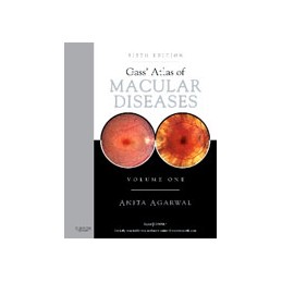
Autor: Anita Agarwal
2-Volume Set - Expert Consult: Online and Print