• Zamawiaj do paczkomatu
  • Płać wygodnie

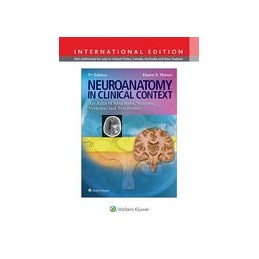
Neuroanatomy in Clinical Context

An Atlas of Structures, Sections, Systems, and Syndromes

9781469832029
242,55 zł
218,29 zł Zniżka 24,26 zł Brutto
Najniższa cena w okresie 30 dni przed promocją: 218,29 zł
Ilość
Produkt niedostępny
Nakład wyczerpany (niedostępny u wydawcy)

  Dostawa

Wybierz Paczkomat Inpost, Orlen Paczkę, DPD, Pocztę, email (dla ebooków). Kliknij po więcej

  Płatność

Zapłać szybkim przelewem, kartą płatniczą lub za pobraniem. Kliknij po więcej szczegółów

  Zwroty

Jeżeli jesteś konsumentem możesz zwrócić towar w ciągu 14 dni*. Kliknij po więcej szczegółów

Opis
Neuroanatomy in Clinical Context , Ninth Edition provides everything the student needs to master the anatomy of the central nervous system, all in a clinical setting. Clear explanations;
abundant MRI, CT, MRA, and MRV images; full-color photographs and illustrations; hundreds of review questions; and supplemental online resources combine to provide a sound anatomical base for integrating neurobiological and clinical concepts. In thus applying neuroanatomy clinically, the atlas ensures student preparedness for exams and for rotations. This authoritative approach—combined with such salutary features as full-color stained sections, extensive cranial nerve cross-referencing, and systems neurobiology coverage—sustains the legacy of this revolutionary teaching and learning tool as the neuroanatomy atlas.
New and hallmark features elucidate neuroanatomy and systems neurobiology for course success!
  • NEW! Chapter on Herniation Syndromes decodes the elegant relationship between brain injury and resulting deficit.
  • NEW! Clinical information integrated throughout the text is screened in blue for quick identification on the page.
  • NEW! Enhanced clinical images emphasize clarity and detail like never before, including full-color images replacing many in black and white, higher-resolution brain scans, and reprocessed spinal cord and brainstem images.
  • MRIs complement full-color anatomical illustrations, allowing for visualization of structures both as they appear to the unaided eye and on imaging studies.
  • Unique, full-color illustrations integrate clinical images of representative lesions with the corresponding deficits highlighted.
  • Full-color stained sections facilitate the easy identification of anatomical features.
  • Dozens of pathway drawings superimposed over MRIs connect structure with function of neural pathways.
Located on thePoint, this atlas’s companion website offers a variety of supplemental
learning resources to maximize study and review time!
  • Question bank featuring over 280 USMLE-style and chapter-review style questions
  • Bonus dissection photographs and brain slice series
Szczegóły produktu
LWW
54968
9781469832029
9781469832029

Opis

Rok wydania
2014
Numer wydania
9
Oprawa
miękka foliowana
Liczba stron
368
Wymiary (mm)
5806 x 7742
Waga (g)
1157
Komentarze (0)